
How peptides work depends on their amino acid sequence, three-dimensional shape and ability to interact with a particular biological target. Many peptides act as signalling molecules. They bind to receptors on the surface of a cell and trigger a chain of events inside it, while others interact with enzymes, ion channels or proteins involved in cellular regulation.
This selectivity allows different peptides to participate in processes such as metabolism, immune signalling, tissue maintenance, growth and communication between cells. However, identifying an interaction is only the first step in understanding peptide biological activity. Researchers must also determine whether the interaction activates or blocks a target, which pathway follows, how strong the response is and how long the signal lasts.
Peptide behaviour is therefore best understood as a sequence: molecular structure influences target binding, target binding changes cellular signalling, and cellular signalling produces a measurable biological response.
What Are Peptides?
Peptides are molecules formed from amino acids joined by peptide bonds. A peptide may contain only a few amino acids or a much longer sequence.
There is no single universal length boundary between peptides vs proteins, although peptides are generally shorter and structurally less complex.
The order of amino acids is important because each amino acid has distinct chemical properties. Some carry an electrical charge, some interact readily with water, and others favour hydrophobic environments. Their arrangement influences how the chain folds, which targets it can recognise and how stable it remains under biological or experimental conditions.
Naturally occurring peptides, including neuropeptides studied in research, can function as hormones, neurotransmitters, growth signals, immune mediators and local regulators. Peptides can also be synthesised in laboratories so that researchers can examine a natural sequence, test an analogue or investigate how a structural change alters activity.
Peptides, Amino Acids and Proteins
Although these terms are related, they describe different levels of molecular organisation.
| Feature | Amino acid | Peptide | Protein |
| Basic structure | A single organic building unit | A chain of amino acids joined by peptide bonds | One or more longer amino acid chains with an organised structure |
| Typical complexity | Low | Variable, but usually lower than a protein | Often includes complex folding and multiple functional regions |
| Common role | Used to build peptides and proteins | Frequently involved in signalling, regulation or molecular recognition | Performs structural, catalytic, transport and regulatory functions |
| Experimental focus | Metabolism, synthesis and incorporation | Receptor binding, signalling, stability and biological activity | Folding, enzyme activity, interactions and cellular function |
Calling peptides the building blocks of proteins can be misleading. Amino acids are the building blocks, while peptides are amino acid chains that may have their own independent biological functions.
Quick Answer: How Do Peptides Work?
Peptides work by interacting with compatible molecular targets. In many cases, a peptide binds to a receptor on the cell surface. This binding changes the receptor’s shape or activity, allowing it to pass a signal into the cell. The signal may then activate enzymes, produce second messengers, alter ion movement or change the expression of particular genes.
The process can be summarised in six stages:
- The peptide adopts a structure influenced by its amino acid sequence.
- It encounters a compatible receptor or molecular target.
- Binding activates, blocks or modifies the target.
- The target initiates a peptide signalling pathway.
- The cell produces a measurable response.
- The signal ends when the peptide is removed, degraded or released from the target.
This general model explains many peptide mechanisms, but not every peptide follows exactly the same route. The result depends on the peptide, the target, the cell type and the surrounding conditions.
How Peptides Work: From Structure to Biological Response
Amino Acid Sequence Determines Structure
A peptide’s sequence determines more than its length. It influences charge, solubility, flexibility and the tendency to form structural features such as turns, loops or helices. These properties affect whether the peptide can remain stable, reach a target and fit the target’s binding region.
Even a single amino acid substitution may change activity. It can strengthen or weaken binding, alter selectivity, make the molecule more vulnerable to enzymatic breakdown or produce a different three-dimensional conformation. This relationship between structure and function is a central part of peptide science.
Peptides Recognise Particular Targets
Molecular recognition depends on complementary shape and chemistry. A peptide and its target interact through forces such as hydrogen bonding, electrostatic attraction, hydrophobic interactions and van der Waals forces.
The strength of the interaction is described as binding affinity. Selectivity refers to how strongly the peptide favours one target over other possible targets. High affinity does not automatically mean perfect selectivity, and binding alone does not prove that a meaningful response will follow.
Binding Changes Target Activity
After binding, a peptide may stabilise an active or inactive form of its target. An activating peptide can cause a receptor to recruit intracellular proteins or switch on an enzyme domain. An inhibitory peptide may occupy the binding site without producing the normal signal or may interfere with an interaction needed for activation.
Some effects occur rapidly, within seconds or minutes. Others take longer because the initial signal must alter gene transcription, protein production or cell behaviour.
Intracellular Signals Carry the Message
Cell-surface receptors allow peptides to influence the cell without physically entering it. When a receptor is activated, internal signalling proteins relay the message through a series of controlled molecular events.
These events often involve phosphorylation, in which enzymes add phosphate groups to proteins and change their activity. Signals may also be carried by small intracellular molecules known as second messengers. A pathway can amplify the original interaction, meaning that a limited number of occupied receptors can influence many molecules inside the cell.
Cells Produce a Measurable Response
The final response depends on which proteins and pathways are present in the cell. Possible outcomes include changes in enzyme activity, ion movement, secretion, metabolism, cell migration, gene expression, proliferation or survival.
The same receptor can sometimes produce different responses in different cell types. Each cell contains its own combination of signalling proteins, regulatory enzymes and genes, so receptor activation is interpreted within that cellular context.
The Signal Is Terminated
Biological signals must be controlled. A response may end when the peptide separates from the receptor, is degraded by peptidases or is removed from the surrounding environment. The receptor may also be desensitised, chemically modified or pulled into the cell through internalisation.
Inside the cell, enzymes can remove phosphate groups, break down second messengers and return signalling proteins to their inactive states. Signal termination prevents continuous stimulation and helps cells remain responsive to later changes.
Major Peptide Receptors and Molecular Targets
Many peptides have limited ability to cross lipid cell membranes because they are relatively large and often contain charged or hydrophilic groups. As a result, cell-surface receptors are major targets in research on peptide signalling and receptor interactions. Other peptides act on extracellular enzymes, membrane channels or protein interaction surfaces.
G Protein-Coupled Receptors
G protein-coupled receptors, or GPCRs, span the cell membrane and transmit information by activating intracellular G proteins. When a peptide binds, the receptor changes conformation and affects enzymes or ion channels inside the cell.
Depending on the receptor and G protein involved, this can increase or decrease cyclic adenosine monophosphate, activate phospholipase C or alter intracellular calcium. GPCR signalling is highly regulated and may produce different effects in different tissues.
Enzyme-Linked Receptors
Some peptide signals bind to receptors that possess enzyme activity or are closely associated with enzymes. Receptor tyrosine kinases are an important example. Ligand binding can bring receptor units together and trigger phosphorylation on the intracellular portion of the receptor.
The phosphorylated sites then recruit signalling proteins, allowing pathways such as PI3K-Akt and MAPK-ERK to become active. In IGF-1 LR3 research, these pathways are examined for their influence on metabolism, protein synthesis, growth and other cellular functions.
Ion Channels and Membrane Proteins
Certain peptides interact directly or indirectly with ion channels. They may increase or decrease the movement of calcium, sodium, potassium or chloride across a membrane. Changes in ion flow can alter electrical activity, secretion, contraction and communication between cells.
Not every peptide that affects an ion channel binds to the channel itself. Some first activate a receptor, with downstream signalling then modifying channel behaviour.
Enzymes and Protein-Protein Interactions
Peptides can bind to enzymes and change their catalytic activity. They may also reproduce a short interaction region from a larger protein and compete for a protein-binding surface.
This makes peptides useful tools for investigating protein-protein interactions that may be difficult to study with smaller molecules. These interactions often cover broad or relatively flat surfaces, and a flexible peptide may contact several important points across the interface.
Important Peptide Signalling Pathways
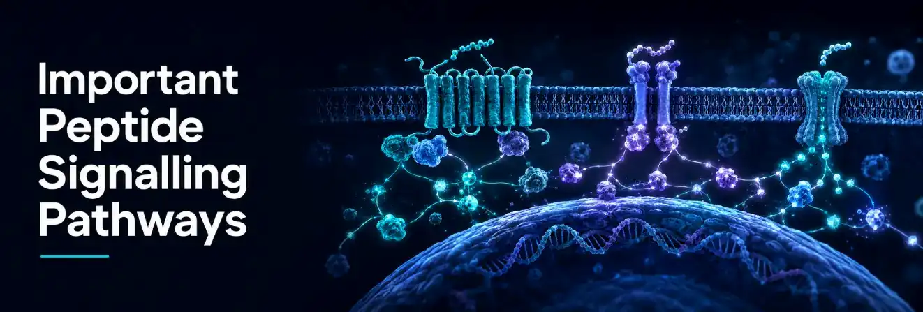
Peptide pathways are networks rather than isolated switches. A receptor may activate several branches, and different pathways can influence one another. The following systems are commonly examined, but their involvement must be established for each peptide and model.
| Signalling pathway | Typical initiating event | Common experimental readouts |
| cAMP-PKA | Activation or inhibition of adenylyl cyclase through a GPCR | cAMP levels, PKA activity and phosphorylation of downstream proteins |
| PLC–IP3–DAG–Calcium | GPCR or receptor activation stimulates phospholipase C | Intracellular calcium, protein kinase C activity and second-messenger levels |
| PI3K-Akt-mTOR | Activation of an enzyme-linked receptor or associated signalling complex | Akt or mTOR phosphorylation, glucose-related responses and protein synthesis markers |
| MAPK-ERK | Sequential activation of kinase proteins | ERK phosphorylation, gene expression, proliferation or differentiation markers |
| JAK-STAT | Receptor-associated Janus kinases phosphorylate STAT proteins | STAT phosphorylation, nuclear movement and target-gene expression |
cAMP and Protein Kinase A
Some GPCRs regulate adenylyl cyclase, the enzyme that produces cyclic AMP. Cyclic AMP activates protein kinase A and other effectors, which then modify downstream proteins. The pathway can influence metabolism, secretion, ion-channel activity and gene transcription.
Because cAMP can be measured with sensitive assays, it is often used to determine whether a test peptide activates or inhibits a particular GPCR.
PLC, IP3, DAG and Calcium
Phospholipase C splits a membrane lipid into IP3 and DAG signalling. IP3 promotes the release of calcium from internal cellular stores, while DAG contributes to protein kinase C activation.
Calcium is a versatile intracellular signal involved in secretion, contraction, enzyme control and gene regulation. Researchers may therefore monitor calcium changes as an early indicator of receptor activation.
PI3K-Akt-mTOR
The PI3K-Akt-mTOR network integrates signals related to nutrient availability, metabolism, protein synthesis, growth and cell survival. Activation usually involves a sequence of membrane recruitment and phosphorylation events.
Detection of Akt or mTOR phosphorylation can show that a pathway has responded, but it does not by itself establish the complete biological outcome. Timing, cell type and activation of parallel pathways must also be considered.
MAPK-ERK
The MAPK-ERK cascade passes information through a series of protein kinases. It can respond to signals associated with growth, stress, differentiation and cell movement.
The duration of ERK activation may matter as much as its intensity. A short pulse and sustained activation can lead to different cellular outcomes, which is why time-course experiments are valuable.
JAK-STAT
In JAK-STAT signalling, receptor-associated Janus kinases phosphorylate STAT proteins. Activated STATs can move into the nucleus and regulate gene expression.
This pathway is commonly associated with cytokine and growth-related signalling. Not all peptides activate JAK-STAT, so pathway involvement should be demonstrated rather than assumed from a general biological effect.
Peptide Agonists, Antagonists and Modulators
Receptor binding can produce several functional outcomes. Classifying a peptide by its effect provides more information than binding affinity alone.
Agonists
An agonist binds to a receptor and promotes an active state. A full agonist can produce the maximum response available in a particular experimental system when sufficient receptors are occupied.
The measured maximum still depends on the assay. A peptide may appear highly active in one cell line and less active in another because receptor density and downstream signalling capacity differ.
Antagonists
An antagonist binds without activating the receptor and reduces the effect of an agonist. A competitive antagonist occupies the same or an overlapping binding site, while other antagonists may bind elsewhere and alter receptor behaviour.
Antagonists are useful for investigating peptide mechanisms because they can help determine whether a measured response depends on a proposed receptor.
Partial Agonists and Biased Signalling
A partial agonist activates a receptor but produces a lower maximum response than a full agonist in the same system. Some ligands also favour one signalling branch over another, a phenomenon known as biased signalling.
This means receptor activation is not always a simple on-or-off event. The conformation stabilised by a bound peptide can influence which intracellular proteins are recruited and which responses dominate.
What Determines Peptide Biological Activity?
Peptide biological activity is shaped by the molecule, its target and the experimental environment. An observed experimental response cannot be attributed to the peptide sequence alone without considering these other variables.
Key factors include:
- Sequence and conformation:These determine whether the peptide can adopt a target-compatible shape.
- Affinity:Stronger binding may allow activity at lower experimental concentrations, but does not guarantee a stronger final response.
- Selectivity:Interaction with additional receptors or enzymes can produce unexpected readouts.
- Concentration and exposure time:Responses may change in size or character as exposure conditions change.
- Receptor expression:Cells with few or no compatible receptors may show little response.
- Pathway availability:Different cells contain different signalling proteins and feedback systems.
- Solubility and aggregation:Poorly dissolved or aggregated material may reduce the available concentration and distort results.
- Chemical and enzymatic stability:Breakdown can shorten exposure or create fragments with different properties.
- Purity and identity:Impurities, incorrect sequences or degradation products can affect an assay.
- Temperature, pH and handling:Experimental conditions can alter peptide structure and stability.
These variables explain why two studies using apparently similar peptides may not produce identical results.
Natural, Synthetic and Modified Peptides
Naturally Occurring Peptides
Natural peptides are produced by organisms through controlled biological processes. Many begin as parts of larger precursor proteins and are released by enzymatic cleavage. Further processing may be required before the mature peptide becomes active.
Natural function provides a useful starting point when studying naturally occurring peptides such as MOTS-C, but isolating a peptide from its normal biological context can change its apparent behaviour. Concentration, location, release timing and interaction with binding proteins all contribute to its activity in a living system.
Synthetic Peptides
Chemical peptide synthesis allows researchers to produce defined peptide sequences, including synthetic research compounds such as Semax. Synthetic material can be used to test receptor binding, map an active region or compare sequence variants under controlled conditions.
Synthetic does not automatically mean identical in every practical respect to a naturally processed peptide. Folding, terminal modifications, disulfide bonds, purity and analytical confirmation remain important.
Modified Peptide Analogues
Researchers may replace amino acids, modify the peptide backbone, protect the ends of the chain or introduce cyclisation. These changes can alter affinity, selectivity, solubility, conformation and resistance to enzymatic degradation.
Modification may improve one property while weakening another. Greater stability, for example, does not necessarily preserve receptor activity. Each analogue requires direct testing rather than assumptions based on the original sequence.
Large sets of sequence variants are often compared using peptide libraries.
How Peptides Are Studied in Laboratory Research

Research peptides are examined in laboratory settings through complementary methods. No single assay can establish binding, mechanism and biological significance at the same time.
Analytical Confirmation
Before functional testing, researchers may assess identity, purity and molecular mass. Chromatographic and mass-spectrometric methods help determine whether the sample contains the expected sequence and whether significant impurities or degradation products are present.
This step matters because an unexpected assay result may reflect sample quality rather than the intended peptide mechanism.
Receptor-Binding Assays
Binding assays measure whether a peptide interacts with a proposed target and estimate properties such as affinity or competition with another ligand. These experiments are useful for confirming molecular recognition.
Binding does not necessarily demonstrate activation. Functional assays are required to show what the interaction does to the receptor or cell.
Cell-Based Functional Assays
Cells expressing a target receptor can be used to measure second messengers, calcium movement, enzyme activity, protein phosphorylation, reporter-gene expression or other responses.
Controls help distinguish a target-dependent effect from background variation. These may include untreated cells, a known reference ligand, a receptor antagonist or cells that do not express the proposed target.
Concentration-Response and Time-Course Experiments
A concentration-response experiment measures how the observed effect changes across a defined range of experimental concentrations. It can help estimate potency and maximum response within that assay system.
A time-course experiment shows when the response begins, reaches its peak and declines. Combining these approaches gives a clearer picture than measuring a single concentration at one time point.
Pathway and Biomarker Analysis
Researchers can examine phosphorylation, gene expression, protein abundance, secretion or metabolic changes to determine which pathways respond. Using pathway inhibitors, receptor-blocking tools or genetic methods can provide stronger evidence for causation.
A biomarker should not be interpreted in isolation. Changes in one downstream molecule may result from more than one upstream pathway.
In Vitro and In Vivo Models
In vitro systems include purified proteins, isolated membranes, cultured cells and tissue preparations. They provide control over experimental variables and can reveal detailed peptide mechanisms.
In vivo models incorporate absorption, distribution, metabolism, excretion and interactions between tissues. They can answer broader biological questions but introduce more variables. Findings from either type of model require interpretation within its limits.
All research-use-only peptides should be handled according to applicable institutional procedures, safety requirements and research-use restrictions. Laboratory findings should not be interpreted as guidance for personal or clinical use.
Batch-specific results are reported in each Certificate of Analysis. For guidance on batch identity and supporting documentation, see our Research Peptides Australia guide.
Challenges When Interpreting Peptide Research
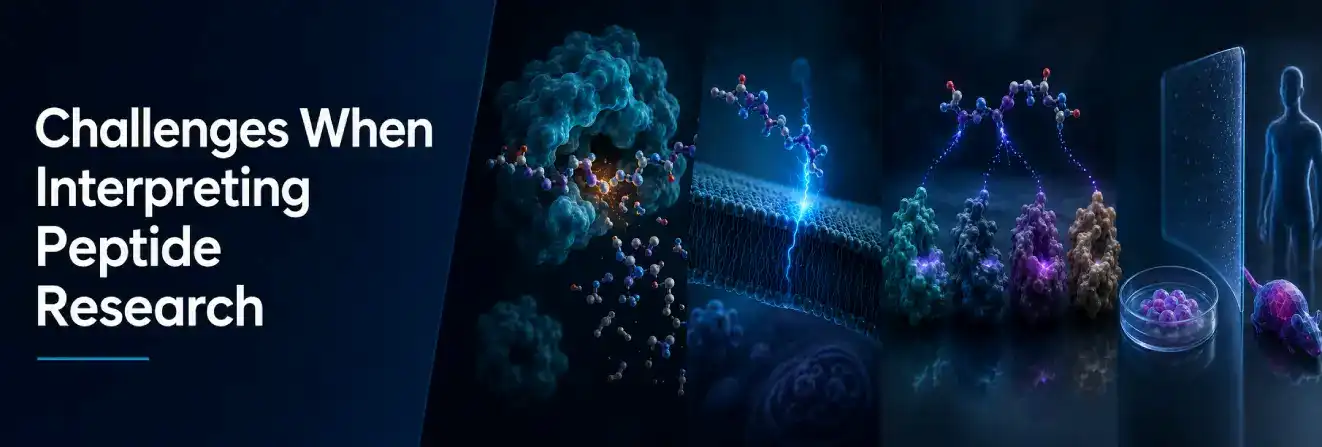
Peptide research can produce convincing molecular data while still leaving important biological questions unresolved.
Stability and Degradation
Peptide bonds can be broken by proteolytic enzymes. Oxidation, deamidation, hydrolysis or aggregation may also change a sample during preparation or storage. If stability is not monitored, the actual exposure may differ from the intended one.
Membrane Permeability
Many peptides do not readily cross cell membranes. An effect attributed to an intracellular target therefore requires evidence that the peptide entered the cell or reached that target through an appropriate delivery method.
Off-Target Activity
A peptide may interact with more than one receptor, particularly at higher experimental concentrations. Similar receptor families can also share structural features. Receptor-blocking experiments and selectivity panels help test whether the proposed target explains the result.
Model-Specific Responses
Cell lines can differ in receptor number, signalling proteins and metabolism. Animal species may also process a peptide differently or express receptors in different patterns. These differences can influence both the strength and type of response.
Limits of Translating Laboratory Findings
Findings from biochemical assays, cultured cells or animal models should be interpreted within the limits of each experimental system. Laboratory studies can identify molecular interactions, signalling pathways and plausible biological mechanisms, but results obtained in one model should not automatically be extrapolated to another without appropriate validation.
This distinction is particularly important when preliminary findings are promoted beyond what the underlying evidence can support.
Peptides Compared With Proteins and Small Molecules
Peptides differ from both small molecules and larger proteins in size, structure, target interactions and experimental characteristics.
| Property | Peptides | Proteins | Small molecules |
| Relative size | Small to medium amino acid chains | Usually larger and more structurally complex | Generally much smaller |
| Common targets | Cell-surface receptors, enzymes and protein interfaces | Receptors, ligands and broad protein surfaces | Enzyme pockets, receptors and intracellular targets |
| Target recognition | Often high because several residues contact the target | Often highly specific | Variable depending on structure |
| Membrane penetration | Commonly limited | Usually very limited without transport mechanisms | Often better, although highly variable |
| Biological stability | Frequently limited by enzymatic degradation | Variable and dependent on structure | Often more resistant to proteolysis |
| Research considerations | Sequence, folding, purity, solubility and degradation | Folding, modifications, aggregation and expression system | Solubility, metabolism, selectivity and off-target binding |
These differences influence experimental design. A method suitable for a stable small molecule may not account for peptide adsorption, proteolysis or structural sensitivity.
Explore our research peptide shop to browse compounds supplied for laboratory research.
How Peptides Work in Research:
Understanding how peptides work requires more than identifying a peptide and listing its reported effects. The process begins with amino acid sequence and molecular structure, continues through selective interaction with a receptor or other target, and produces a response through connected signalling pathways.
The observed activity depends on receptor expression, pathway availability, exposure conditions, peptide stability and sample quality. Binding results, cellular responses and whole-organism findings therefore answer different questions and should not be treated as interchangeable evidence.
Well-designed peptide research combines analytical confirmation, receptor studies, functional assays, suitable controls and careful interpretation. This approach connects molecular interaction with biological response while recognising the limits of each experimental model.
FAQs About How Peptides Work
How Do Peptides Work in Laboratory Research?
Peptides can interact with receptors, enzymes, ion channels or other molecular targets in experimental systems. Researchers measure the resulting changes in signalling pathways, enzyme activity, gene expression or other cellular responses to investigate peptide mechanisms.
How Do Peptides Interact With Receptors?
Peptide-receptor interactions depend on molecular shape, amino acid sequence and chemical compatibility. Binding may activate, inhibit or modify receptor activity, which can then influence intracellular signalling pathways and measurable experimental responses.
What Determines Peptide Activity in an Experimental Model?
Peptide activity depends on factors such as sequence, structure, target affinity, receptor expression, concentration, stability and experimental conditions. Results may therefore differ between cell types, assay systems and research models.
How Quickly Can Peptide Signalling Be Detected in Research?
The timing depends on the peptide, molecular target and experimental readout. Some receptor-signalling events can be detected within seconds or minutes, while downstream changes in gene expression or cellular behaviour may require longer observation periods.