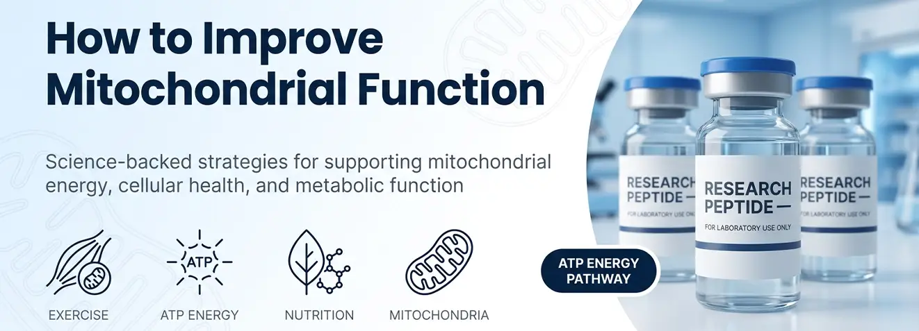
How to improve mitochondrial function is one of the most actively researched questions in modern biology, and the answer is clear: mitochondrial performance is substantially modifiable through lifestyle.
Exercise, nutrition, sleep quality, thermal exposure, and certain investigational compounds all demonstrably alter mitochondrial dynamics, with the strongest evidence pointing to structured physical activity as the primary driver.
Mitochondria do more than produce cellular energy. They also participate in apoptosis, calcium signalling, reactive oxygen species regulation, and the synthesis of several important metabolites.
This article covers each strategy in depth, drawing on peer-reviewed evidence structured for both readability and scientific precision.
What Are Mitochondria and Why Does Their Performance Matter?
Mitochondria are double-membraned organelles found in nearly every nucleated cell in the body.
Their primary role is to produce adenosine triphosphate (ATP) through oxidative phosphorylation, a process carried out along the inner mitochondrial membrane by the electron transport chain (ETC). These mechanisms are explained in more detail in our guide to mitochondrial function.
Beyond energy production, mitochondria also:
- Regulate reactive oxygen species (ROS) as intracellular signalling molecules
- Buffer calcium to modulate muscle contraction, neurotransmitter release, and gene expression
- Initiate apoptosis when cells are irreparably damaged
- Participate in steroid hormone and haem biosynthesis
- Encode 13 essential proteins within their own genome, mitochondrial DNA (mtDNA)
Cells with high energy demands—including neurons, cardiomyocytes, skeletal muscle fibres, and hepatocytes—typically contain abundant mitochondria and may be especially affected by impaired mitochondrial function. Mitochondrial dysfunction has been associated with type 2 diabetes, neurodegenerative disorders, cardiovascular disease, and sarcopenia.
What Causes Mitochondrial Dysfunction?
Mitochondrial dysfunction can be inherited, but the most common cause is secondary impairment driven by modifiable lifestyle and environmental factors, including cellular ageing, making prevention and reversal genuinely achievable.
Identifying these causes is the starting point for any effective strategy to support mitochondrial function.
| Cause | Mechanism | Modifiable? |
| Chronic oxidative stress | Excess ROS oxidises mtDNA, ETC proteins, and membrane lipids | Yes, diet, exercise, sleep |
| Physical inactivity | Reduces PGC-1α signalling, suppressing mitochondrial biogenesis | Yes, structured exercise |
| Poor sleep quality | Disrupts circadian control of mitochondrial fusion, fission, and mitophagy | Yes, sleep hygiene |
| Nutrient deficiencies | CoQ10, B vitamins, magnesium, and iron are all ETC cofactors | Yes, dietary improvement |
| Chronic systemic inflammation | Pro-inflammatory cytokines impair membrane potential and biogenesis | Yes, lifestyle, nutrition |
| High refined carbohydrate intake | Promotes glycation of mitochondrial proteins; elevates ROS output | Yes, dietary choices |
| Environmental toxin exposure | Heavy metals and persistent organics directly inhibit ETC complexes | Partially |
| Biological ageing | Accumulation of somatic mtDNA mutations; declining NAD+ concentrations | Partially; lifestyle slows decline |
How to Improve Mitochondrial Function: Six Evidence-Based Strategies
The following strategies are supported by published research and represent the strongest available evidence for improving or maintaining healthy mitochondria. They are ordered from most to least established in terms of human clinical data.
1. Exercise and Mitochondria: the Most Potent Biogenesis Signal
Exercise is the most potent activator of PGC-1α (peroxisome proliferator-activated receptor gamma coactivator 1-alpha), the master driver of mitochondrial biogenesis, the process by which cells form new mitochondria.
Different exercise modalities target mitochondria through distinct pathways:
| Exercise Type | Primary Mitochondrial Mechanism | Key Research Finding |
| High-Intensity Interval Training (HIIT) | Amplifies AMPK and PGC-1α signalling via repeated metabolic stress cycles | Robinson et al. (2017, Cell Metabolism): HIIT increased mitochondrial protein synthesis more than moderate continuous exercise, particularly in older adults |
| Moderate aerobic exercise (walking, cycling, swimming) | Sustained PGC-1α activation; increases mitochondrial volume density in slow-twitch muscle | 150+ minutes/week associated with measurably higher mitochondrial density in observational studies |
| Resistance training | Increases mitochondrial biogenesis in fast-twitch fibres, a population largely unaddressed by aerobic protocols | Additive effects when combined with aerobic training on mitochondrial volume density |
| Zone 2 (low-intensity steady-state) | Preferentially trains mitochondrial fat oxidation; improves respiratory efficiency. | Commonly used in longevity-focused research protocols; improves metabolic flexibility. |
For comprehensive mitochondrial stimulus: 2–3 HIIT sessions, 150+ minutes of moderate aerobic activity, and 2 resistance sessions per week.
- What Is the Best Nutrition for Mitochondria?
Nutrition supports mitochondrial function in two main ways: macronutrients provide substrates for the Krebs cycle and the electron transport chain, while micronutrients act as cofactors in energy-producing reactions.
Dietary patterns with the strongest mitochondrial evidence:
| Pattern | Mitochondrial Benefit |
| Mediterranean | Polyphenols and omega-3s preserve membrane integrity and reduce oxidative damage. |
| Ketogenic/low-carb | Shifts fuel to fat oxidation; ketones yield more ATP per unit of ROS than glucose. |
| 16:8 intermittent fasting | Triggers mitophagy; preserves intracellular NAD+ concentrations |
Key polyphenols that activate PGC-1α and SIRT1:
| Compound | Source | Mechanism |
| Resveratrol | Red grapes, berries, peanuts | SIRT1 activation; supports muscle mitochondrial biogenesis |
| Quercetin | Capers, apples, red onions | Inhibits NLRP3 inflammasome; reduces mitochondrial oxidative damage |
| EGCG | Green tea | Activates AMPK; improves mitochondrial uncoupling |
| Sulforaphane | Broccoli, Brussels sprouts | Activates Nrf2; upregulates mitochondrial antioxidant defences |
Essential micronutrients for healthy mitochondria:
| Nutrient | Role in ETC / ATP Synthesis | Dietary Sources |
| CoQ10 | Electron shuttle between Complexes I/II and III; membrane antioxidant | Organ meats, oily fish, nuts, spinach |
| NAD+ (NR/NMN) | Primary electron acceptor in Krebs cycle; declines ~50% by mid-life | Dairy, beef, mushrooms, fish |
| Magnesium | Cofactor for ATP synthase and 300+ enzymatic reactions | Pumpkin seeds, leafy greens, dark chocolate |
| B vitamins (B1–B5) | Precursors to NADH and FADH2 — primary ETC electron donors | Whole grains, eggs, legumes, meat |
| Iron | Structural component of cytochrome proteins (Complexes II, III, IV) | Red meat, legumes, shellfish |
| Alpha-lipoic acid | Cofactor in pyruvate dehydrogenase complex; regenerates vitamins C and E | Spinach, broccoli, organ meats |
| L-Carnitine | Transports fatty acids across inner mitochondrial membrane for beta-oxidation | Red meat, poultry, dairy, fish |
| Selenium | Component of glutathione peroxidase; protects mitochondrial membranes | Brazil nuts, tuna, eggs |
3. Sleep and Mitochondrial Health: The Overlooked Recovery Window
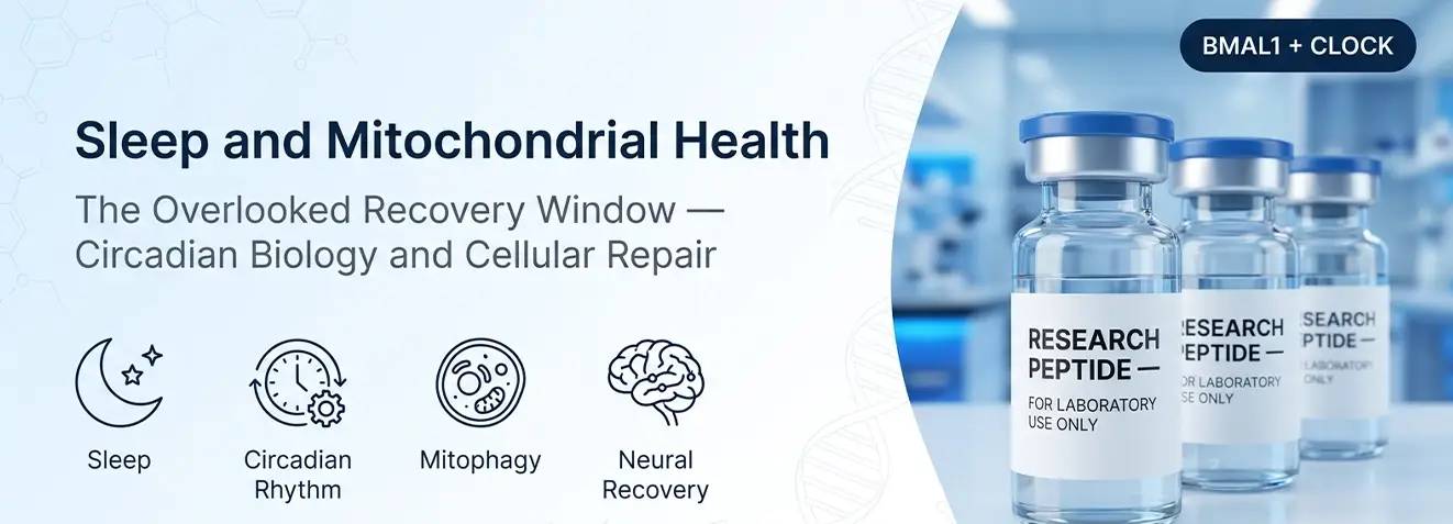
Sleep and mitochondrial health are closely linked through circadian biology. Core clock proteins, including BMAL1 and CLOCK, help regulate daily patterns in mitochondrial metabolism and quality-control processes such as fusion, fission, biogenesis, and mitophagy. These pathways are discussed further in our guide to neuropeptide signalling and mitochondrial regulation.
When these rhythms are disrupted, mitochondrial quality control deteriorates. Research consequences of poor sleep include:
- Increased ROS production in brain tissue, particularly in the prefrontal cortex and hippocampus
- Impaired mitophagy, resulting in accumulation of damaged mitochondria that would normally be cleared
- Reduced ATP output in neurons, cells that cannot easily dilute damaged organelles through division
- Accelerated mtDNA oxidative damage in sleep-deprived animal models
- Suppression of the glymphatic system, which clears metabolic waste (including oxidised mitochondrial proteins) from the brain during slow-wave sleep
- Can Heat and Cold Exposure Improve Mitochondrial Adaptation?
Temperature manipulation has emerged as a practical tool for stimulating mitochondrial adaptation, with mechanistic evidence across both ends of the thermal spectrum.
Heat exposure
A 2018 study published in the Journal of Applied Physiology demonstrated that repeated heat application to cultured human skeletal muscle cells without any exercise increased mitochondrial number and enhanced respiratory capacity. The mechanism involves heat shock proteins (HSPs), which stabilise ETC membrane proteins, assist in the refolding of oxidatively damaged components, and upregulate biogenesis signalling via HSF1 activation.
Regular sauna use (4–7 sessions per week at 80–100°C) is associated in longitudinal Finnish cohort data with:
- Reduced all-cause mortality
- Improved cardiovascular markers including blood pressure and arterial stiffness
- Decreased circulating levels of C-reactive protein (CRP) and other inflammatory markers
Cold exposure
Controlled cold exposure can activate brown adipose tissue (BAT), which is rich in mitochondria and specialised for non-shivering thermogenesis. However, the magnitude of this response varies between individuals and exposure protocols.
Key mitochondrial effects include:
- Upregulation of mitochondrial uncoupling protein 1 (UCP1), which increases mitochondrial volume and metabolic rate in BAT
- Enhanced glucose and lipid uptake in BAT and skeletal muscle
- Reduction of ROS accumulation in peripheral tissues due to metabolic slowing during cold application
- When combined with exercise, amplification of PGC-1α-mediated mitochondrial biogenesis signalling in skeletal muscle
- How Does Oxidative Stress Affect the Mitochondrial Membrane?
Mitochondria are both the primary source and target of reactive oxygen species (ROS), the drivers of oxidative stress at the cellular level.
Under normal conditions, ROS act as signalling molecules, but when antioxidant capacity is exceeded, oxidative stress damages mtDNA, ETC proteins, and the inner mitochondrial membrane.
Practical strategies to reduce this burden include:
- Limit alcohol intake:
ethanol metabolism generates excess NADH, disrupting the NAD+/NADH ratio and increasing mitochondrial ROS output
- Avoid tobacco smoke:
acrolein and other components directly inhibit ETC Complex I
- Reduce visceral adiposity:
adipose tissue, particularly visceral fat, secretes pro-inflammatory adipokines that impair mitochondrial biogenesis signals
- Prioritise dietary antioxidants:
vitamins C and E, selenium, and zinc support glutathione peroxidase and superoxide dismutase (SOD), the primary enzymatic defences of the mitochondrial membrane
- Manage chronic psychological stress:
sustained cortisol elevation activates NADPH oxidase and promotes systemic inflammation, directly impairing mitochondrial membrane potential
- How Do Peptides Support Mitochondrial Energy? Investigational Compounds
Emerging peptide research has identified several compounds with potential mitochondrial effects.
| Compound | Mechanism Under Investigation | Research Stage |
| MOTS-C | Mitochondria-derived peptide encoded in the 12S rRNA gene of mtDNA; activates AMPK, improves insulin sensitivity, and promotes mitochondrial biogenesis in skeletal muscle; may counteract age-associated mitochondrial decline. | Preclinical (animal models); early human observation |
| Humanin | Encoded within the 16S rRNA gene of mtDNA; cytoprotective against apoptosis in neurons and cardiomyocytes; preclinical evidence for reduced oxidative stress and preserved mitochondrial membrane integrity under metabolic challenge. | Preclinical; human correlation studies |
| NAD+ | Restore intracellular NAD+ concentrations, which decline ~50% by mid-life; NAD+ is the primary electron carrier in oxidative phosphorylation and substrate for sirtuin-mediated mitochondrial regulation. | Phase I/II human clinical trials; published data on skeletal muscle mitochondrial markers |
| Epitalon | Tetrapeptide (Ala-Glu-Asp-Gly) studied for effects on telomere length and mitochondrial membrane lipid peroxidation; some preclinical literature suggests reduced oxidative damage to cardiolipin in the inner mitochondrial membrane. | Preclinical; limited human data |
| BPC-157 | Synthetic pentadecapeptide studied for cytoprotective and anti-inflammatory effects; preclinical research suggests modulation of nitric oxide signalling, which intersects with mitochondrial electron transport and ROS management | Preclinical only |
Mitochondrial Health Tips at a Glance

The following table consolidates the key mitochondrial health tips covered in this article, with primary mechanisms and evidence grading:
| Strategy | Primary Mitochondrial Mechanism | Evidence Grade |
| HIIT + Resistance Training | PGC-1α-driven biogenesis; increased mitochondrial volume density | Strong (RCTs) |
| Zone 2 aerobic training | Improves fat oxidation efficiency; metabolic flexibility | Strong |
| Mediterranean / whole-food diet | Polyphenol-driven SIRT1/PGC-1α activation; reduced oxidative load | Moderate–Strong |
| 16:8 intermittent fasting | Mitophagy induction; NAD+ preservation | Moderate (human) |
| 7–9 hours quality sleep | Circadian-regulated mitochondrial dynamics; glymphatic clearance | Moderate–Strong |
| Sauna (4–7x/week) | HSP upregulation; increased biogenesis without exercise | Moderate (cohort) |
| Cold water immersion | BAT activation; UCP1 expression; ROS reduction | Moderate (preclinical + human) |
| CoQ10 supplementation | Direct ETC electron shuttle; membrane antioxidant | Moderate (clinical) |
| NAD+ precursors (NR/NMN) | Restores electron carrier availability; sirtuin activation | Early clinical trials |
| MOTS-c (research only) | AMPK activation; skeletal muscle biogenesis | Preclinical only |
Recognising Reduced Mitochondrial Energy Output
Mitochondrial dysfunction rarely presents as a single identifiable symptom. More commonly, it appears as a cluster of overlapping signs that accumulate over time. The following are not diagnostic criteria, but they represent patterns frequently observed in research literature on mitochondrial impairment:
- Persistent fatigue that does not resolve with rest or normal sleep
- Exercise intolerance or disproportionate fatigue following physical activity
- Cognitive slowing, reduced processing speed, difficulty with working memory or concentration
- Muscle weakness or cramping at low exertion levels
- Difficulty maintaining stable blood glucose between meals
- Slow recovery from illness, surgery, or physical stress
- Heightened sensitivity to environmental stressors including light, temperature changes, and noise
These presentations overlap with many other conditions; clinical evaluation by a qualified practitioner is required before drawing any conclusions about mitochondrial status.
The Research Case for Mitochondrial Health
Mitochondrial function sits at the intersection of energy, ageing, and cellular resilience.
The evidence reviewed here indicates that exercise, nutrition, and sleep are the most accessible and best-supported lifestyle approaches for maintaining mitochondrial health. Evidence for thermal exposure is promising but less established.
Emerging investigational compounds add a further dimension to this research landscape, though their application remains strictly preclinical.
For research scientists and curious minds alike, understanding how to improve mitochondrial function is less about any single intervention and more about the cumulative effect of evidence-based choices made consistently over time.
Frequently Asked Questions
How can I repair my mitochondria naturally?
Regular exercise, a nutrient-rich diet, quality sleep, and stress management can support healthy mitochondrial function over time.
What vitamins increase mitochondria?
B vitamins help support normal mitochondrial energy production, but no vitamin alone increases mitochondria.
What damages mitochondria the most?
Physical inactivity, smoking, poor diet, chronic sleep deprivation and excessive alcohol are among the biggest contributors to mitochondrial dysfunction.
What are the symptoms of weak mitochondria?
Reduced mitochondrial function may be associated with fatigue, muscle weakness, exercise intolerance and difficulty concentrating.
What foods rebuild mitochondria?
No food rebuilds mitochondria directly, but whole foods rich in vegetables, fruits, legumes, nuts and oily fish support mitochondrial health.
How long does it take to restore your mitochondria?
Healthy lifestyle changes can improve mitochondrial function gradually over several weeks to months.
What foods are bad for mitochondria?
Ultra-processed foods, excess added sugar, and excessive alcohol may negatively affect mitochondrial function.
Does CoQ10 repair mitochondria?
CoQ10 supports mitochondrial energy production but has not been proven to repair mitochondria.
Which organ has a lot of mitochondria?
The heart contains one of the highest concentrations of mitochondria because of its constant energy demand.
Sources:
https://nigms.nih.gov/biobeat/2022/11/science-snippet-atps-amazing-power
https://www.ncbi.nlm.nih.gov/books/NBK9896/