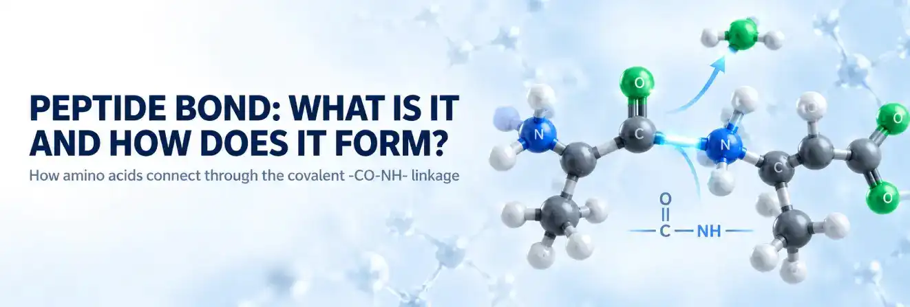
A peptide bond is a covalent chemical bond that connects one amino acid to another, creating the backbone of peptides and proteins. It forms between the carboxyl group of one amino acid and the amino group of another, producing the characteristic amide linkage represented as –CO–NH–.
This apparently simple connection has important structural consequences. Peptide bonds do more than hold amino acids together: their geometry, resonance and restricted rotation help determine how a polypeptide chain can move and fold.
Understanding peptide bond formation therefore provides a foundation for understanding peptide chemistry, protein structure, biological synthesis and the processes that break peptides and proteins back into smaller components.
Peptide Bond at a Glance
| Question | Answer |
|---|---|
| What is a peptide bond? | A covalent amide bond linking amino acid residues. |
| Where does it form? | Between the carbonyl carbon of one residue and the nitrogen of the next. |
| Basic structure | –C(=O)–NH– |
| Bond type | Covalent |
| Geometry | Approximately planar |
| Rotation | Restricted around the peptide C–N bond |
| Common configuration | Trans |
| How is it broken? | Hydrolysis |
What Is a Peptide Bond?
A peptide bond is an amide-type covalent bond that joins adjacent amino acid residues in a peptide or protein chain. More specifically, the bond forms between the carbonyl carbon associated with the carboxyl group of one amino acid and the nitrogen associated with the amino group of the next amino acid.
The resulting connection can be represented in simplified form as:
–C(=O)–NH–
Once amino acids have been incorporated into a peptide chain, they are commonly described as amino acid residues because chemical groups involved in the joining process are no longer present in exactly the same form as in the free amino acids.
It is classified as an amide linkage between amino acids, formed when the amino group of one amino acid is joined to the carboxyl group of another. Repetition of this linkage along a chain creates the characteristic peptide backbone.
The peptide bond should not be confused with the many weaker interactions that can occur after a protein folds. The backbone connection itself is covalent, whereas hydrogen bonding, ionic interactions and hydrophobic interactions can help organise that backbone into a three-dimensional structure.
Where Does the Peptide Bond Form Between Amino Acids?
A typical amino acid contains an α-amino group, an α-carboxyl group, a hydrogen atom and a side chain attached to the α-carbon.
During peptide bond formation, the groups involved are:
- the α-carboxyl groupof one amino acid; and
- the α-amino groupof another amino acid.
The carbonyl carbon of the first residue ultimately becomes bonded to the nitrogen of the second.
A simplified representation is:
Amino acid 1–COOH + H₂N–Amino acid 2 → Amino acid 1–CO–NH–Amino acid 2 + H₂O
This basic reaction explains the peptide bond in amino acid chemistry and shows why the finished linkage contains both a carbonyl group and nitrogen.
The amino acid side chains, often represented as R groups, are not normally the groups forming the standard backbone peptide bond. They remain attached to their respective α-carbons and contribute many of the chemical differences between individual amino acids.
Is the Linkage Covalent?
Yes. A peptide bond is a covalent bond.
Covalent bonds involve the sharing of electrons between atoms and are generally much stronger than non-covalent interactions such as hydrogen bonds.
This distinction is important in proteins. Peptide bonds create the durable covalent backbone of a polypeptide, while weaker interactions between different regions of the chain help determine how that backbone folds.
How Does Peptide Bond Formation Occur?
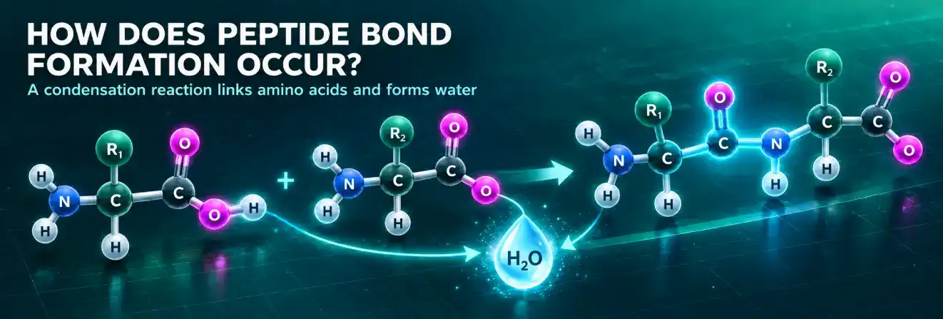
Peptide bond formation can be described at a basic chemical level as a condensation reaction between amino acid groups.
In the simplified representation, components corresponding to a hydroxyl group from the carboxyl side and a hydrogen from the amino side are removed, corresponding to the formation of water. A new carbon–nitrogen connection then links the two amino acid residues.
This process is commonly called a condensation reaction or dehydration reaction when peptide formation is introduced in basic chemistry and biochemistry.
The Condensation Reaction Between Two Amino Acids
The process can be illustrated using two free amino acids.
One provides the carboxyl group:
–COOH
The other provides the amino group:
–NH₂
The simplified overall change can be written as:
–COOH + H₂N– → –CO–NH– + H₂O
The newly created –CO–NH– unit is the peptide linkage.
When two amino acid residues become connected in this way, the resulting molecule is called a dipeptide.
Peptide chains can continue to grow as additional amino acid residues are joined through additional peptide bonds.
Peptide Bond Formation Step by Step
At a simplified chemical level, peptide bond formation can be understood in the following sequence:
- Two amino acids are positioned so that the carboxyl functionality of one is associated with the amino functionality of the other.
- The carboxyl side contributes components corresponding to a hydroxyl group.
- The amino side contributes a hydrogen.
- These components correspond to the formation of a molecule of water in the overall condensation equation.
- The carbonyl carbon becomes covalently linked to nitrogen.
- The new –CO–NH– unit is the peptide bond.
- The joined amino acids are now residues within a dipeptide.
This model clearly shows the chemical relationship between amino acids, water and the resulting amide linkage.
In cells, peptide bond formation occurs through specialised biological machinery rather than through this simplified model alone.
Watch how peptide bonds form between amino acids.
Why Is Water Associated With Peptide Bond Formation?
Water appears in the overall condensation equation because the atoms removed from the participating groups correspond to H₂O.
The carboxyl side contributes OH, while the amino side contributes H in the simplified model. Together, these account for one water molecule.
For this reason, educational diagrams often describe peptide bond formation as a dehydration reaction.
This representation is useful for understanding the relationship between the structures before and after bond formation. It should not be interpreted as a complete description of every intermediate step involved in biological protein synthesis.
From Dipeptides to Longer Peptide Chains
One peptide bond connecting two amino acid residues produces a dipeptide.
Adding additional residues creates progressively longer chains. Terms such as tripeptide can be used for specific small chains, including the sequence found in GHK-Cu peptide, while peptide and polypeptide describe longer amino acid sequences.
There is no universally sharp chemical boundary in the peptides vs proteins distinction at which every peptide suddenly becomes a protein.
The terms are influenced by factors such as chain length, molecular size, structure and biological context.
The important structural principle remains the same: amino acid residues in research peptides and other polypeptide chains are connected sequentially through peptide bonds to create the polypeptide backbone.
Peptide Bond Structure and Chemical Properties
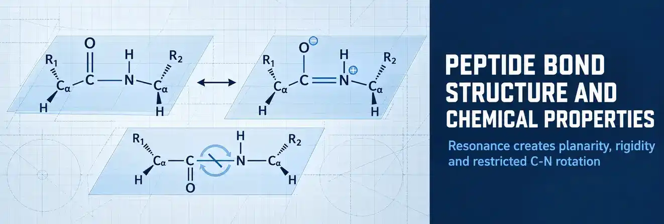
The peptide bond structure is more complex than a simple single C–N bond might suggest.
The central peptide group contains a carbonyl carbon, carbonyl oxygen, nitrogen and hydrogen or substituted nitrogen group. Electron delocalisation across this region gives the peptide linkage properties that influence the shape and flexibility of peptide and protein chains.
| Feature | Peptide Bond |
| Bond type | Covalent amide bond |
| Basic linkage | –CO–NH– |
| Forms between | Carboxyl functionality and amino functionality of adjacent residues |
| Overall introductory reaction | Condensation |
| Molecule represented as released | H₂O |
| C–N rotation | Restricted |
| Geometry | Approximately planar |
| Common arrangement | Predominantly trans |
| Main location | Peptide and protein backbones |
What Does the Linkage Look Like?
The central chemical pattern is:
C(=O)–N
For a common backbone peptide linkage, it may be represented more fully as:
–C(=O)–NH–
The carbonyl carbon is double-bonded to oxygen and bonded to nitrogen. The α-carbons of the neighbouring amino acid residues lie on either side of this peptide unit.
This arrangement repeats throughout a typical polypeptide chain:
–N–Cα–C(=O)–N–Cα–C(=O)–N–
Side chains extend from the α-carbons, while the repeating backbone contains the peptide bonds.
Resonance and Partial Double-Bond Character
One of the most important properties of the peptide bond structure is resonance, which gives the C–N linkage partial double-bond character and restricts free rotation.
Electrons are not completely localised in a way that would make the carbonyl C=O bond and neighbouring C–N bond behave as entirely independent bonds. Instead, electron delocalisation gives the C–N connection some double-bond character.
As a result, the peptide C–N bond behaves differently from an ordinary freely rotating carbon–nitrogen single bond.
This partial double-bond character contributes directly to the rigidity of the peptide unit.
Why Is the Linkage Planar and Rigid?
Restricted rotation around the peptide C–N bond means that the atoms of the peptide group tend to remain in a relatively fixed planar arrangement.
The relationship can be summarised as:
Resonance → partial double-bond character → restricted C–N rotation → planar peptide unit
This rigidity is important because a protein backbone is not a completely flexible chain in which every bond rotates freely.
Instead, relatively rigid peptide units are connected through bonds around the α-carbon that have substantially greater rotational freedom. The combination of rigid and rotatable regions helps define the conformations available to a polypeptide.
Cis vs Trans Peptide Bonds
A peptide bond can be described in terms of cis and trans configurations based on the relative positioning of neighbouring groups around the peptide linkage.
The trans configuration is strongly favoured for most peptide bonds because it generally places adjacent α-carbons and their attached groups farther apart, reducing steric crowding.
Cis peptide bonds occur less frequently.
Peptide bonds involving proline are notable because the energetic difference between cis and trans configurations is smaller than it is for many other residues. This makes proline a structurally distinctive residue in short sequences such as KPV peptide (Lys-Pro-Val). As a result, cis configurations are more relevant when proline is involved, although trans arrangements remain common.
The possibility of cis–trans isomerisation matters because changing the peptide bond configuration can alter the geometry of the polypeptide backbone.
Peptide Bonds in Proteins and Polypeptides
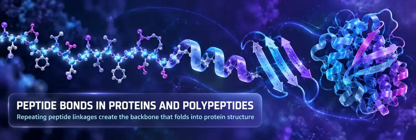
The peptide bond performs the same fundamental chemical role across peptides and proteins, although the differences between peptides and proteins extend beyond this shared backbone linkage.
When repeated many times, these bonds form the protein’s primary backbone.
The identity and order of the side chains attached to that backbone define the amino acid sequence, while the structural characteristics of the backbone influence how the chain can fold.
How Peptide Bonds Build the Protein Backbone
A polypeptide can be viewed as a repeating sequence of backbone units connected by peptide bonds.
Each amino acid residue contributes part of the repeating structural pattern. The side chain attached to each α-carbon differs between amino acids, but the backbone arrangement follows the same general framework.
This organisation gives proteins two important levels of chemical information:
Backbone: a recurring structure connected by peptide bonds.
Side chains: chemically diverse groups that influence charge, polarity, hydrophobicity, interactions and folding.
The peptide backbone therefore provides continuity, while the side-chain sequence creates much of the chemical diversity.
N-Terminus and C-Terminus
A linear peptide or polypeptide has directionality.
At one end is the N-terminus, associated with a free amino group. At the other end is the C-terminus, associated with a free carboxyl group.
By convention, amino acid sequences are usually written from the N-terminus toward the C-terminus.
The residues between these ends are linked through peptide bonds, giving the chain a defined chemical direction rather than making it a symmetrical polymer.
Why Peptide Bond Geometry Matters for Protein Structure
The rigidity of the peptide bond limits one type of backbone movement, but neighbouring single bonds remain capable of rotation.
Two important backbone angles are commonly called phi (φ) and psi (ψ).
Phi describes rotation around the N–Cα bond, while psi describes rotation around the Cα–carbonyl carbon bond.
Not every theoretical combination of these angles is physically possible because atoms cannot occupy the same space. Steric effects therefore restrict the conformations available to a polypeptide chain.
Protein folding is consequently shaped by both chemical bonding and three-dimensional geometry.
Peptide Bonds and Secondary Protein Structure
Peptide bonds also provide chemical groups that participate in hydrogen bonding.
The carbonyl oxygen can act as a hydrogen-bond acceptor, while the N–H group of many peptide linkages can participate as a hydrogen-bond donor.
Repeated hydrogen bonding between different backbone regions contributes to common forms of protein secondary structure, including:
- α-helices, in which hydrogen bonding helps stabilise a coiled backbone arrangement;
- β-sheets, in which hydrogen bonding occurs between neighbouring or separated stretches of polypeptide backbone.
The hydrogen bonds are not peptide bonds themselves. Peptide bonds form the covalent backbone, while hydrogen bonds help organise that backbone into higher levels of structure.
Explore more research information from Australia Peptide Sciences.
How Are Peptide Bonds Formed in Living Cells?
The simple condensation equation is valuable for learning peptide chemistry, but cells use a more organised process to build proteins.
Protein synthesis occurs on ribosomes, using messenger RNA as the sequence template and transfer RNAs to deliver amino acids.
The chemistry still produces peptide bonds between amino acid residues, but the reactants are activated and positioned by biological machinery.
Peptide Bond Formation on the Ribosome
During translation, amino acids are carried by transfer RNA molecules, commonly abbreviated tRNA.
During translation, the ribosome positions tRNAs carrying specific amino acids so that peptide bonds form between them as the growing polypeptide chain is extended.
The result is transfer of the growing chain to the incoming amino acid, creating a new peptide bond and extending the polypeptide.
The ribosome then continues through the messenger RNA sequence so that further amino acids can be added in the encoded order.
This repeated cycle produces a directional polypeptide chain whose amino acid sequence reflects the genetic information carried by the mRNA.
Chemical Formation vs Biological Peptide Synthesis
The distinction between the introductory chemical model and cellular synthesis is useful:
| Feature | Simplified Condensation Model | Cellular Protein Synthesis |
| Main purpose | Explains the overall chemical relationship | Builds biological polypeptides |
| Amino acids represented as | Free amino acids | Amino acids carried by tRNA |
| Main concept | Condensation and amide formation | Ribosome-mediated peptide transfer |
| Key machinery | Not shown | Ribosome, tRNA and mRNA |
| Product | New peptide bond | Extended polypeptide chain |
| Level of detail | Introductory chemical model | Biological molecular process |
The simplified condensation equation therefore explains what changes chemically, while ribosomal synthesis explains how cells organise and drive the process.
Peptide bonds can also occur through non-ribosomal peptide biosynthesis, where specialised enzymatic machinery assembles peptide products outside the conventional ribosomal pathway. Not every biologically produced peptide is assembled through the same pathway as a conventional ribosomally translated protein.
How Are Peptide Bonds Broken?
Peptide bond formation can be reversed through hydrolysis.
Hydrolysis involves the addition of water across the bond, ultimately separating residues that had been connected through the peptide linkage.
Although peptide bonds can undergo hydrolysis chemically, cleavage under biological conditions is commonly accelerated by specialised enzymes.
Hydrolysis of the Linkage
Hydrolysis is often presented as the conceptual reverse of condensation:
Formation: amino acid residues become linked.
Hydrolysis: a peptide linkage is cleaved through a reaction involving water.
The overall relationship can be summarised as follows:
| Process | Peptide Bond Formation | Peptide Bond Hydrolysis |
| Main effect | Joins residues | Separates residues |
| Water in overall representation | Produced in simplified condensation equation | Consumed during cleavage |
| Peptide bond | Formed | Broken |
| Biological relevance | Peptide and protein synthesis | Peptide and protein breakdown |
A peptide bond is relatively stable under ordinary physiological conditions when no suitable catalyst is present. Biological systems therefore rely on enzymes when controlled peptide-bond cleavage needs to occur efficiently.
Proteases and Peptidases
Enzymes that catalyse peptide-bond hydrolysis include proteases and peptidases.
Different enzymes recognise different substrates or cleavage environments. Some act on large proteins, while others act on shorter peptides or specific regions of a chain.
Controlled peptide-bond cleavage is important in processes such as protein turnover, digestion, activation of precursor proteins and regulation of biological pathways.
The key chemical event remains the same: cleavage of the amide linkage connecting amino acid residues.
Why Are Peptide Bonds Important?
Peptide bonds are fundamental to peptide and protein chemistry because they provide the covalent framework that keeps amino acid sequences connected across many peptide classes, including cellular and recovery research peptides.
Their importance extends beyond simple linkage:
- They connect individual amino acid residues into defined sequences, including longer research peptide sequences such as BPC-157 peptide.
- They create the repeating backbone of peptides and proteins.
- Their resonance limits rotation around the peptide C–N bond.
- Their planar geometry constrains possible backbone conformations.
- Their carbonyl and N–H groups can participate in hydrogen bonding.
- They contribute indirectly to the formation of α-helices, β-sheets and other protein structures.
- Their controlled formation allows cells to assemble polypeptides.
- Their controlled hydrolysis allows biological systems to process and degrade peptides and proteins.
The combination of strength and structural restriction is particularly important. A peptide backbone must remain covalently connected while still having enough flexibility around neighbouring bonds to adopt functional three-dimensional structures.
For laboratory materials designated as research-use-only peptides, understanding peptide chemistry does not establish safety, effectiveness or suitability for personal or clinical use. Such materials should be handled according to applicable institutional procedures, safety requirements and research-use restrictions.
Browse available research products in the peptide shop.
Peptide Bond vs Other Bonds in Proteins
Proteins contain several different types of bonds and interactions, and they should not be treated as interchangeable.
A peptide bond forms the backbone by linking consecutive amino acid residues. Other connections can stabilise or organise a protein after that backbone has formed.
| Bond or Interaction | Main Role | Covalent? | Typical Location |
| Peptide bond | Connects consecutive amino acid residues | Yes | Polypeptide backbone |
| Disulfide bond | Can stabilise folded structures or connect chain regions | Yes | Between cysteine side chains |
| Hydrogen bond | Helps stabilise secondary and tertiary structure | No | Backbone and/or side chains |
| Isopeptide bond | Creates an alternative amide linkage | Yes | Specific amino acid functional groups |
Peptide Bond vs Hydrogen Bond
A peptide bond is a covalent connection between amino acid residues. A hydrogen bond is a weaker non-covalent interaction involving suitable hydrogen donors and acceptors.
Both matter to proteins, but for different reasons.
Peptide bonds maintain the continuous amino acid backbone. Hydrogen bonds help organise portions of that backbone and can also occur between side chains.
For example, hydrogen bonding between peptide backbone groups contributes to α-helices and β-sheets without replacing or breaking the peptide bonds themselves.
Peptide Bond vs Disulfide Bond
A disulfide bond is a covalent connection that can form between the sulfur-containing side chains of two cysteine residues.
Unlike a peptide bond, it does not normally create the sequential backbone connection between one residue and the next.
Instead, a disulfide bond can connect regions that may be separated in the amino acid sequence. This can help stabilise a folded protein or link different polypeptide chains.
Therefore:
Peptide bond: builds the primary backbone.
Disulfide bond: can crosslink cysteine-containing regions.
Peptide Bond vs Isopeptide Bond
A conventional peptide bond joins the α-carboxyl group of one amino acid residue with the α-amino group of another.
An isopeptide bond is also an amide linkage, but it involves at least one functional group outside this standard backbone arrangement, such as an amino or carboxyl group located in an amino acid side chain.
The distinction is important because saying that amino acids are connected by an amide bond does not automatically mean that the linkage is a standard backbone peptide bond.
Understanding Peptide Bonds in Amino Acids and Proteins
The peptide bond is the covalent link that joins amino acids into peptides, polypeptides and proteins. It forms between the carbonyl carbon of one residue and the nitrogen of the next, creating the repeating –CO–NH– backbone.
Resonance gives the C–N bond partial double-bond character, restricting rotation and helping maintain a planar structure, while neighbouring bonds provide flexibility for folding.
In cells, ribosomes and tRNA coordinate peptide bond formation during protein synthesis. Hydrolysis reverses this process, and proteases or peptidases can cleave peptide bonds when biological systems need to process or break down peptides and proteins in a controlled manner.
FAQs About Peptide Bonds
What is a peptide bond in simple terms?
A peptide bond is the covalent link that connects one amino acid to another. It forms between the carbonyl carbon of one amino acid and the nitrogen of the next, helping create the backbone of peptides and proteins.
How can you identify a peptide bond?
A peptide bond can be identified by finding the –C(=O)–N– group in the molecular structure. The key connection is the bond between a carbonyl carbon and the nitrogen of the neighbouring amino acid residue.
Where are peptide bonds found?
Peptide bonds occur throughout the backbone of peptides, polypeptides and proteins. They join amino acid residues together in a continuous sequence.
How strong is a peptide bond?
A peptide bond is a stable covalent amide bond. Resonance gives the C–N bond partial double-bond character, which makes it relatively rigid and limits free rotation around the linkage.
What is the difference between a peptide and a peptide bond?
A peptide is a molecule made from amino acids linked in a chain, while a peptide bond is the specific chemical connection that joins those amino acids together.
What is the difference between a peptide and a protein?
Peptides and proteins are both built from amino acids connected by peptide bonds. Peptides are generally shorter chains, while proteins are usually larger and more structurally complex, often folding into defined three-dimensional shapes.
How many peptide bonds are in a peptide?
In a linear peptide containing n amino acid residues, there are generally n − 1 peptide bonds. For example, a dipeptide contains one peptide bond, a tripeptide contains two, and a peptide with 10 amino acid residues contains nine peptide bonds.
Is a peptide bond polar?
Yes. The peptide group is polar because the carbonyl oxygen and amide nitrogen create an uneven distribution of electron density and a molecular dipole. However, the peptide linkage itself is generally uncharged under physiological conditions. Its polarity also allows peptide backbone groups to participate in hydrogen bonding.