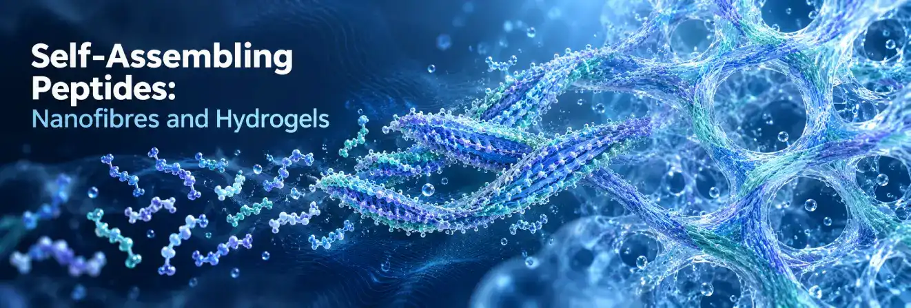
Self-assembling peptides are amino acid sequences that organise into larger structures through interactions between neighbouring molecules. Depending on their sequence and environment, they can form nanofibres, nanotubes, nanoribbons and water-rich networks known as hydrogels.
These materials are studied in supramolecular chemistry, biomaterials science and molecular engineering. Their structures link peptide composition with measurable properties, including network stiffness, molecular transport and nanoscale organisation.
Understanding these relationships helps researchers interpret experiments involving peptide hydrogels, functional nanomaterials and three-dimensional cell culture models.
What Are Self-Assembling Peptides?
Self-assembling peptides form supramolecular structures under suitable physicochemical conditions. Many assemblies are stabilised predominantly by non-covalent forces rather than new covalent bonds between their building blocks. Individual peptides may therefore associate into structures far larger than the original molecules.
What Makes a Peptide Self-Assembling?
The peptide sequence determines the distribution of hydrophobic, polar and charged side chains. For example, alternating hydrophobic and hydrophilic residues may create distinct molecular surfaces that favour regular packing. Self-assembly becomes thermodynamically favourable when the overall free-energy balance supports association, although assembly kinetics can determine which structures appear experimentally.
Not every peptide forms an ordered nanomaterial: some remain dispersed, while others produce irregular aggregates.
Sequence, molecular conformation and surrounding conditions must all be considered. Researchers distinguish ordered assembly from precipitation by combining observations of molecular conformation, morphology and material behaviour. This avoids treating any visible solid or cloudy suspension as evidence of successful self-assembly.
Peptide Self-Assembly vs Peptide Synthesis
Peptide synthesis creates a defined amino acid sequence by forming covalent peptide bonds; solid-phase synthesis is widely used in research laboratories. Peptide self-assembly is the subsequent association of peptide molecules through intermolecular forces, without necessarily forming additional covalent bonds.
A successfully synthesised peptide is not necessarily self-assembling. Likewise, visible aggregation does not establish the presence of ordered nanofibres or a hydrogel. Those conclusions require experimental characterisation.
How Does Peptide Self-Assembly Work?
Self-assembly depends on molecular interactions, peptide conformation and solvent conditions. Thermodynamics influences which structures are energetically favourable, while kinetics controls the rate and pathway of their formation.
Non-Covalent Interactions Driving Peptide Self-Assembly
Several forces commonly operate together:
- Hydrogen bonding:Backbone and side-chain interactions stabilise secondary structures and ordered assemblies.
- Hydrophobic interactions:Non-polar regions associate to reduce unfavourable exposure to water.
- Electrostatic interactions:Opposite charges can attract, while like charges may repel.
- Van der Waals interactions:Numerous weak atomic contacts contribute to molecular packing.
- Aromatic interactions:Aromatic groups, particularly phenylalanine residues, can influence assembly through interactions that may include π–π stacking.
For example, hydrogen bonding can support β-sheet organisation, while hydrophobic interactions encourage neighbouring molecular surfaces to associate. Their relative contributions depend on sequence and experimental conditions.
Solvent molecules also play an active role: changes in hydration can favour or destabilise arrangements that otherwise appear similar.
The strength of a single molecular interaction cannot reliably predict the final structure without considering the full assembly environment.
The Role of Secondary Structures
β-Sheets contain hydrogen-bonded peptide strands and commonly support fibrillar or ribbon-like assemblies. α-Helices can create amphipathic surfaces that associate into coiled coils. β-Hairpins, containing two strands joined by a turn, can undergo conformational changes that favour fibrillar networks.
Secondary structure influences molecular packing but does not uniquely determine morphology. Similar motifs can produce different assemblies when charge distribution, chemical modifications or solution conditions change.
From Individual Peptides to Supramolecular Structures
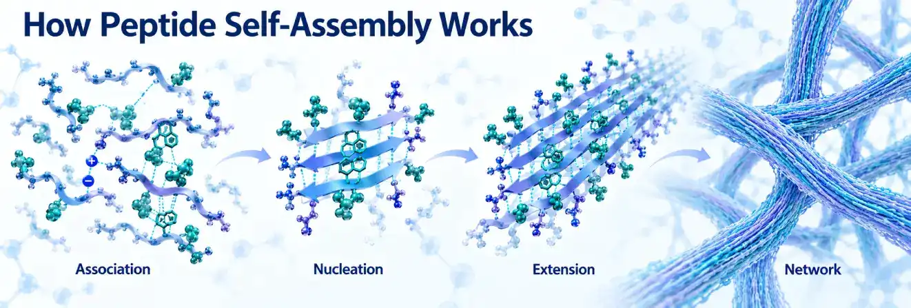
A common assembly pathway involves four stages:
- Association:Peptides establish favourable intermolecular contacts.
- Nucleation:Small ordered clusters may initiate further growth.
- Extension:Additional molecules join growing fibres or other assemblies.
- Hierarchical organisation:Primary structures associate into bundles or interconnected networks.
This is not a universal sequence. Competing pathways and kinetic trapping can generate structures that are not the most thermodynamically stable arrangement. Consequently, samples with identical compositions may differ after different preparation histories.
Main Types of Self-Assembling Peptides
Peptide families are often grouped by molecular design and characteristic assembly behaviour.
Ionic-Complementary Peptides
Ionic-complementary peptides combine patterned charged residues with hydrophobic regions. Electrostatic interactions, hydrophobic association and backbone hydrogen bonding influence their organisation.
EAK16, containing alanine, glutamic acid and lysine, and RADA16, containing arginine, alanine and aspartic acid, are established research examples. Both have been investigated for β-sheet-rich nanofibres; RADA16 is also widely studied in hydrogel networks.
Peptide Amphiphiles
Peptide amphiphiles contain regions with different affinities for water. Some join a peptide segment to a hydrophobic alkyl chain, while others obtain amphiphilic character from their amino acid arrangement. In aqueous systems, association of hydrophobic regions can produce nanofibres, micelles or ribbons, depending on molecular geometry and conditions.
Aromatic Dipeptides and Short Peptides
Diphenylalanine (FF) consists of two phenylalanine residues and is a common model for investigating aromatic interactions and molecular packing.
FF-based systems can form nanotubes, fibrils or crystalline structures under different conditions. Fmoc-FF, an N-terminally protected derivative, is well studied for supramolecular hydrogel formation. Chemical modifications can alter assembly behaviour. Separately, researchers have investigated copper-mediated co-assembly of GHK, the copper-binding peptide found in GHK-Cu complexes, with the structure-forming tripeptide FFD to produce a hydrogel.
β-Hairpin and Cyclic Peptides
Designed MAX-family β-hairpin peptides can adopt conformations that favour fibrillar hydrogel networks. Certain cyclic peptides containing alternating D- and L-amino acids stack through backbone hydrogen bonding to form nanotubular structures. Ring geometry and side-chain composition influence their dimensions and surface properties.
| Peptide class | Representative examples | Commonly investigated structures |
| Ionic-complementary | RADA16, EAK16 | β-Sheet-rich nanofibres and hydrogels |
| Peptide amphiphiles | Alkyl-linked peptide amphiphiles | Nanofibres, micelles and ribbons |
| Aromatic short peptides | FF, Fmoc-FF | Nanotubes, fibrils and hydrogels |
| β-Hairpin peptides | MAX-family peptides | Fibrillar hydrogel networks |
| Cyclic peptides | Alternating D/L cyclic peptides | Nanotubular assemblies |
These associations are representative, not guaranteed outcomes for every peptide or laboratory condition. Classification schemes also overlap: an aromatic peptide may be amphiphilic, and an ionic-complementary sequence may form β-sheets. These labels describe useful design features rather than mutually exclusive categories.
Additional educational resources on peptide chemistry and laboratory research are available through Australia Peptide Sciences.
What Factors Influence Peptide Self-Assembly?
Environmental conditions can change molecular charge, solvation, assembly kinetics and the stability of existing structures.
pH and Ionic Strength
pH affects protonation of peptide termini and ionisable residues, including aspartic acid, glutamic acid, lysine and histidine. Altered charge patterns can favour or disrupt particular assemblies. Dissolved ions screen electrostatic interactions, potentially reducing repulsion or changing packing. Increasing ionic strength does not always promote assembly; ion identity and sequence both matter.
Temperature and Peptide Concentration
Temperature influences molecular motion, solvent interactions and transitions between dispersed and assembled states. Concentration affects molecular encounters and network connectivity. At one concentration, nanofibres may remain dispersed; under other conditions, they may form a connected network. More peptide does not necessarily mean a stronger or more homogeneous gel.
Solvent Composition and External Stimuli
Solvent composition changes peptide solvation and the balance of hydrophobic, electrostatic and hydrogen-bonding interactions. Specially engineered systems may also respond to enzymatic modifications, light or redox changes, triggering assembly or disassembly.
Such responsiveness is sequence- and chemistry-dependent. For example, researchers investigated an engineered phosphorylated peptide precursor incorporating the LKKTETQ sequence, identified as TB500 in the study. Enzymatic dephosphorylation of this precursor triggered nanofibre formation and hydrogelation.
| Experimental factor | Potential influence on self-assembly |
| pH | Changes protonation and charge interactions |
| Ionic strength | Alters electrostatic screening and packing |
| Temperature | Influences kinetics and structural transitions |
| Peptide concentration | Affects association and network connectivity |
| Solvent composition | Changes solvation and molecular interactions |
| Enzymes or light | Regulates responsive systems designed for these triggers |
Reproducible comparisons require consistent conditions and appropriate controls. Studies should distinguish a change in assembly rate from a change in the eventual structure. For example, two conditions might produce similar mature fibres but markedly different times to reach that state. Both observations matter when interpreting experimental results.
Peptide Nanofibres: Formation, Structure and Properties
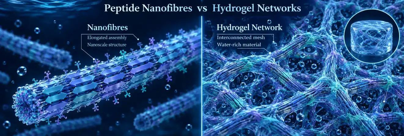
Peptide nanofibres are elongated, organised assemblies whose dimensions and surface chemistry depend on the constituent molecules.
How Do Self-Assembling Peptides Form Nanofibres?
Directional intermolecular association produces fibre growth. In many β-sheet systems, backbone hydrogen bonding aligns peptide strands, while hydrophobic and electrostatic forces stabilise the assembly. Additional molecules extend the structures, which may remain isolated, form bundles or contribute to larger networks. Growth depends on molecular packing and competing assembly pathways.
Structural Properties of Peptide Nanofibres
Fibre diameter, length, flexibility, charge and packing affect interactions with neighbouring fibres and other molecules. Changing the distribution of hydrophobic residues or attaching functional groups can modify surface properties and molecular recognition.
Researchers characterise these features to connect nanoscale morphology with material behaviour. Apparent fibre widths can differ between imaging methods because electron-microscopy preparation and AFM probe geometry influence measurements.
Reporting imaging technique and sample state is therefore as important as quoting a characteristic fibre dimension.
Nanofibres vs Other Peptide Nanostructures
Nanotubes contain a hollow interior; nanoribbons and nanotapes are flatter elongated assemblies. Micelles often segregate hydrophobic and hydrophilic regions, whereas vesicles enclose a compartment within a molecular layer.
These architectures differ in accessible surfaces and internal spaces. Importantly, the presence of nanofibres alone does not prove that a hydrogel has formed.
How Do Self-Assembling Peptide Hydrogels Form?
Self-assembling peptide hydrogels are water-rich materials in which peptide molecules create a sufficiently interconnected three-dimensional network. Many contain entangled or physically associated nanofibres whose collective structure gives rise to measurable bulk properties.
From Peptide Nanofibres to Hydrogel Networks
Peptides may first form fibrils that subsequently connect throughout a sample. Water occupies the spaces around these assemblies, while network connectivity gives the material resistance to deformation over a relevant timescale. Many networks rely on reversible non-covalent interactions; others incorporate chemical crosslinks or additional components.
Nanofibre formation and gelation are separate observations. Microscopy can reveal fibres, but rheology and complementary structural measurements are needed to determine whether they form a mechanically continuous hydrogel.
Network connectivity can arise from fibre entanglement, junctions or other reversible associations. Consequently, gels with similar-looking fibres may exhibit substantially different stiffness and recovery behaviour.
This video shows researchers 3D-printing structures using self-assembling peptide hydrogel materials.
Structural and Mechanical Properties of Peptide Hydrogels
Network organisation, fibre interactions and water content influence hydrogel stiffness, porosity and viscoelasticity. Viscoelastic materials both store and dissipate energy under deformation.
Oscillatory rheology measures the storage modulus (G′), representing elastic response, and the loss modulus (G″), representing viscous response. When G′ exceeds G″ at specified testing conditions, the response is elastic-dominated.
This alone does not establish network architecture or long-term stability. Frequency, temperature, strain and sample history can all affect the measurements. Researchers also consider whether the reported moduli were obtained within the linear viscoelastic region.
Comparisons made at different deformation amplitudes can otherwise be misleading, especially in networks that soften or rearrange under strain.
Shear-Thinning and Self-Healing Behaviour
Some peptide hydrogels are shear-thinning: their apparent viscosity decreases with increasing shear rate. Some also exhibit self-healing, recovering part of their structure or mechanical response after deformation. Reversible molecular associations may support both behaviours, but they are not equivalent. A material can thin under shear without recovering its original properties, so recovery extent and timescale require separate testing.
Peptide-Based Hydrogels vs Conventional Polymer Hydrogels
Both peptide-based hydrogels and conventional polymer hydrogels retain water within three-dimensional networks. Peptide assemblies often exploit sequence-dependent molecular recognition and non-covalent organisation. Conventional polymer networks may use physical associations, chemical crosslinks or both.
Peptide composition offers one means of tuning molecular interactions, while polymer systems offer other approaches to chemistry and processing. Neither class is universally preferable; measured properties determine research suitability.
How Are Self-Assembling Peptides Studied in the Laboratory?
Researchers use complementary methods to examine peptide identity, secondary structure, nanoscale morphology and bulk mechanical behaviour. No single test establishes every aspect of assembly. Spectroscopy may indicate β-sheet enrichment without showing fibre dimensions, while microscopy may reveal fibres without demonstrating a connected, water-rich gel. Confidence increases when independent measurements agree with a proposed structural model.
Spectroscopic Methods for Studying Peptide Structure
Circular dichroism (CD) spectroscopy measures differences in absorption of circularly polarised light and provides information about α-helical, β-sheet and less ordered conformations. Scattering from concentrated or aggregated samples can complicate its spectra.
Fourier-transform infrared (FTIR) spectroscopy measures vibrational absorption; its amide I region is informative about peptide backbone conformation and hydrogen-bonding environments. Overlapping bands and sample conditions affect interpretation. Together, CD and FTIR can support conclusions about conformational changes during assembly.
Microscopy Techniques for Nanostructure Analysis
Atomic force microscopy (AFM) maps sample topography and can reveal fibre dimensions and surface organisation. Transmission electron microscopy (TEM) examines fine structure using electrons transmitted through a specimen, while scanning electron microscopy (SEM) characterises surface morphology.
Microscopy distinguishes fibres, ribbons, tubes and less organised aggregates, but sample preparation matters. Drying, staining or surface deposition can alter apparent morphology. Where suitable, cryogenic imaging and complementary techniques provide evidence closer to hydrated conditions. Even then, interpretation should distinguish features measured directly from structural models inferred from images. Multiple fields of view and independent preparations help prevent unusual particles from being mistaken for a representative morphology.
Rheological Analysis of Peptide Hydrogels
Rheology links supramolecular organisation with mechanical response. Important measurements include:
- Frequency sweeps:Determine how elastic and viscous responses depend on oscillation frequency.
- Amplitude or strain sweeps:Identify the linear viscoelastic region and response to larger deformation.
- Time-dependent measurements:Track network formation or ageing.
- Recovery tests:Evaluate the extent and timescale of mechanical recovery after deformation.
Testing conditions and sample history must accompany reported values. A fibre dispersion and a continuous gel network may have markedly different mechanical responses.
Additional Structural and Analytical Techniques
Small-angle X-ray scattering (SAXS) probes nanoscale dimensions and organisation; wide-angle X-ray scattering (WAXS) provides information about shorter-range molecular packing. Liquid chromatography and mass spectrometry help establish peptide identity, composition and possible impurities, though they do not independently prove supramolecular morphology.
| Technique | Main research purpose | Important limitation |
| CD spectroscopy | Secondary structure | Scattering and solution effects |
| FTIR spectroscopy | Backbone conformation | Overlapping spectral bands |
| AFM | Surface morphology and dimensions | Surface preparation artefacts |
| TEM / SEM | Nanostructure imaging | Specimen preparation effects |
| Rheology | Viscoelastic and network behaviour | Dependence on test conditions |
| SAXS / WAXS | Nanoscale organisation and packing | Model-dependent interpretation |
| Chromatography / mass spectrometry | Identity and composition | Does not establish assembled morphology |
Assessing Assembly Reproducibility and Peptide Quality
Peptide purity, terminal modifications, solvent composition and preparation history may change observed morphology or mechanics. Accordingly, nominally identical samples can behave differently. A 2025 study by Malik and colleagues combined molecular simulations with FTIR, electron microscopy and rheology to examine a designed peptide hydrogel, illustrating the value of evidence from multiple measurement scales.
Reliable comparisons depend on documented conditions, controls and independent reproducibility. Relevant records include peptide batch information, sample age, solution pH, ionic conditions and measurement temperature. These details are especially valuable when apparently conflicting results arise between laboratories.
The research peptide catalogue provides details of individual compounds available for laboratory research.
Laboratory Research Applications of Self-Assembling Peptides
Self-assembling peptides provide controlled experimental platforms for studying molecular interactions, structured aqueous materials and cell–material behaviour. Their laboratory roles must be distinguished from demonstrated clinical performance.
Three-Dimensional Cell Culture and Extracellular Matrix Models
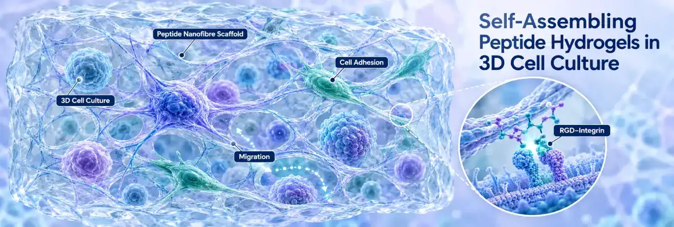
Studies involving cellular research peptides may investigate signalling pathways, while some self-assembling peptide hydrogels provide fibrillar matrices for examining cell adhesion, morphology and migration in three-dimensional culture.
Peptides may incorporate recognition sequences, including arginine–glycine–aspartic acid (RGD), which can interact with certain integrin receptors.
Cellular responses also depend on network stiffness, architecture and cell model. These scaffolds imitate selected extracellular-matrix features, not its complete biological complexity.
The mere inclusion of a cell-binding motif does not guarantee that it remains accessible after assembly. Researchers therefore investigate the combined effect of motif presentation, fibre organisation and matrix mechanics on cultured cells.
Peptide Biomaterials and Tissue Engineering Research
Research scaffolds allow comparisons of amino acid sequence, network mechanics and surface chemistry in laboratory models.
Researchers have also incorporated the KPV motif into longer engineered peptide sequences to investigate self-assembling hydrogel formation.
Studies may examine cell attachment, spatial organisation or responses to different material architectures. Enzyme-responsive motifs can help investigate scaffold changes in controlled experiments.
Findings from these systems do not independently establish safety or effectiveness in living organisms.
Molecular Encapsulation and Controlled Release Studies
Peptide hydrogels are used to examine whether selected molecules remain associated with a network or diffuse through it. Transport depends on molecular size, charge, hydrophobicity and interactions with the peptide matrix. Network organisation can alter diffusion even at similar water contents.
Such experiments clarify transport mechanisms; laboratory release profiles do not establish biological targeting or therapeutic benefit.
Comparisons are particularly informative when they separate passive diffusion from temporary molecular binding or changes in the gel network.
This distinction helps explain why two compounds of similar size may move through the same material at different rates.
Biosensors and Functional Nanomaterials
Nanofibres and nanotubes can provide organised surfaces or templates for electrode modification, molecular recognition and nanoscale material assembly.
Researchers investigate changes in fluorescence, electrical response or analyte binding.
A 2026 review by Wang and colleagues described programmable supramolecular structures, including peptide-based nanopores for molecular detection. These remain experimental platforms requiring validated fabrication and measurements.
What Are the Main Limitations of Self-Assembling Peptide Materials?
Predicting Structure from Peptide Sequence
A sequence can support competing intermolecular interactions, making its final morphology difficult to predict. Small substitutions may change fibre formation, aggregation or gelation.
Researchers can use peptide libraries to compare sequence variants and computational screening to prioritise candidates, but neither approach alone establishes their final assembly morphology.
Many predictions address aggregation tendency rather than fibre dimensions, network formation or rheological properties. These outcomes should not be treated as equivalent endpoints.
Mechanical Stability and Structural Control
Some peptide hydrogels have limited stiffness or change under environmental stress. Increasing concentration or modifying sequence can improve one property while disrupting network homogeneity or molecular accessibility. Evaluation must consider several material characteristics together.
Experimental Reproducibility
Changes in purity, buffer composition, sample preparation and assembly history can alter results. Consistent characterisation and transparent reporting are necessary when comparing studies or batches.
Interpreting Biological Compatibility
Peptide-derived materials cannot automatically be considered biocompatible, biodegradable or non-toxic. Modifications, degradation products and biological interactions matter. Compatibility observed in a cell model does not establish behaviour in more complex systems.
Scalability and Standardisation
Producing larger batches with comparable properties requires control of peptide identity, purity, processing and assembly kinetics. Reproducing the sequence alone does not guarantee consistent supramolecular organisation.
Emerging Directions in Self-Assembling Peptide Research
AI-Assisted Peptide Design and Molecular Simulations
Molecular dynamics simulations help investigate peptide association and packing, while machine-learning systems can prioritise candidate sequences for investigation. A 2025 study by Yang and colleagues used deep learning alongside coarse-grained molecular simulations to examine aggregation propensity in designed decapeptides. However, predicted aggregation is not proof of an ordered hydrogel.
Model assumptions, training data and experimental validation remain central limitations. Predicted structures should be compared with measurements made under comparable solvent and concentration conditions; otherwise, disagreement may reflect the mismatch between simulation assumptions and the laboratory system.
Programmable and Stimuli-Responsive Peptide Materials
Engineered peptides can respond to changes in pH, enzymatic activity, light or redox conditions, sometimes altering assembly reversibly.
Another developing area involves dissipative assemblies, whose organisation depends on continuing energy input or chemical-fuel consumption rather than simply approaching equilibrium. A 2026 review by Schuler and colleagues examined this distinction. Predictable behaviour in complex experimental environments remains an active research challenge.
The Research Value of Self-Assembling Peptides
Self-assembling peptides show how amino acid sequence and molecular interactions can generate nanofibres, hydrogels and other supramolecular architectures. Their value in materials research lies in linking molecular structure to observable morphology, mechanics and transport properties.
Reliable interpretation requires complementary characterisation, controlled experimental conditions and reproducible findings. Computational design is expanding the possibilities, but experimental evidence remains essential.
FAQs About Self-Assembling Peptides
What Is the Self-Assembly of Peptides?
Peptide self-assembly is the organisation of individual peptide molecules into larger structures through intermolecular forces such as hydrogen bonding, hydrophobic interactions and electrostatic attraction. Depending on sequence and conditions, products can include nanofibres, nanotubes or hydrogel networks.
Why Do Some Peptides Self-Assemble?
Their amino acid sequences create chemical surfaces that favour particular intermolecular contacts. Hydrophobic and charged residue patterns, molecular conformation and environmental conditions together determine whether ordered structures form.
What Are Self-Assembling Peptide Hydrogels?
They are water-rich materials containing interconnected peptide assemblies, often nanofibres. Network arrangement and peptide chemistry determine their water retention, mechanical response and stability.
How Are Peptide Nanofibres Formed?
Directional association of peptide molecules produces elongated structures. Hydrogen bonding, hydrophobic forces and electrostatic interactions can stabilise their growth, especially in β-sheet-forming systems.
What Is the Difference Between Peptide Hydrogels and Nanofibres?
A nanofibre is an elongated nanoscale assembly; a hydrogel is a water-containing, mechanically connected network. Nanofibres may form part of a hydrogel, but their presence alone does not demonstrate gelation.
What Conditions Affect Peptide Self-Assembly?
pH, ionic strength, temperature, concentration and solvent composition influence molecular interactions and assembly kinetics. Specially designed systems may also respond to enzymes, light or oxidation conditions.
How Is Peptide Self-Assembly Measured?
CD and FTIR examine conformation; AFM and electron microscopy reveal morphology; SAXS and WAXS probe organisation; and rheology evaluates network mechanics. Multiple methods provide stronger evidence than one measurement.
What Are Self-Assembling Peptides Used for in Research?
Laboratories study them in biomaterials, three-dimensional cell culture, molecular diffusion and functional sensing systems. These experimental applications do not establish clinical effectiveness or suitability for human or veterinary use.
Sources:
https://www.osti.gov/biblio/1820460
https://pmc.ncbi.nlm.nih.gov/articles/PMC4312208/
https://www.nih.gov/news-events/nih-research-matters/injected-liquid-forms-gel-damaged-heart
https://www.energy.gov/science/bes/articles/nature-inspires-new-wave-biotechnology