
Metabolic flexibility is the ability of cells and tissues to adjust their use of carbohydrates and fats in response to changing nutrient availability and energy demands. After feeding, insulin promotes glucose utilisation, while fasting generally increases fatty acid oxidation.
This adaptability depends on mitochondrial function, hormonal signalling and coordinated metabolic pathways. Researchers study its association with insulin resistance and metabolic dysfunction, although causal relationships remain unclear.
Indirect calorimetry and controlled laboratory assessments help measure fuel switching, but no single test provides a universally accepted measure of metabolic flexibility.
How Does Metabolic Flexibility Work?
Fuel selection reflects a coordinated response to substrate supply, transport, storage and cellular ATP requirements. The balance changes during feeding, fasting and increased energy demand rather than switching abruptly between two isolated pathways.
Fuel Utilisation in the Fed State
Following carbohydrate intake, rising blood glucose stimulates insulin secretion from pancreatic beta cells. In skeletal muscle and adipose tissue, insulin signalling promotes translocation of glucose transporter type 4 (GLUT4) to the cell membrane, increasing glucose uptake.
Inside cells, glycolysis converts glucose into pyruvate. Where conditions permit, pyruvate enters mitochondria and is converted to acetyl-coenzyme A (acetyl-CoA), supporting ATP production through the tricarboxylic acid (TCA) cycle and oxidative phosphorylation. Glucose may also be stored as glycogen, particularly in muscle and liver.
Insulin suppresses adipose tissue lipolysis, reducing circulating fatty acid release. Carbohydrate availability also affects enzymes regulating fatty acid oxidation. The fed state therefore tends to favour carbohydrate use without eliminating lipid oxidation.
Fuel Utilisation During Fasting
As insulin concentrations decline, adipose tissue releases more fatty acids through lipolysis. Oxidative tissues can use these fatty acids through mitochondrial beta-oxidation, which produces acetyl-CoA and electron carriers for ATP synthesis.
The liver maintains circulating glucose through glycogenolysis and gluconeogenesis. With longer fasting, hepatic ketogenesis may increase, supplying ketone bodies such as beta-hydroxybutyrate and acetoacetate to other tissues.
Glucose use nevertheless continues. Red blood cells, for example, depend on glycolysis, while other tissues vary the contributions of glucose, fatty acids and ketones. Metabolic flexibility requires coordinating these overlapping demands.
How Energy Demand Changes Fuel Selection
ATP demand is another determinant of substrate choice. During sustained lower-intensity exercise, fatty acids can supply a substantial share of oxidative energy. At higher intensities, carbohydrate utilisation typically rises because glycolysis and carbohydrate oxidation support faster ATP turnover.
The response depends on exercise duration, glycogen availability, mitochondrial capacity and training status. Changes during exercise are informative but are not interchangeable with the metabolic responses to feeding or insulin stimulation.
| Metabolic state | General substrate response | Important regulators |
| Fed | Greater carbohydrate oxidation and nutrient storage | Insulin, GLUT4, PDH |
| Fasting | Greater fatty acid mobilisation and oxidation | Insulin, glucagon, CPT1 |
| Increased energy demand | Variable carbohydrate and fat contributions | AMPK, ATP turnover, mitochondrial enzymes |
These are relative patterns, not exclusive fuel states.
This video explains metabolic flexibility and clarifies common misconceptions about carbohydrate and fat utilisation.
What Regulates Metabolic Flexibility at the Cellular Level?
Cellular adaptation depends on the ability to transport substrates, regulate enzyme activity and match mitochondrial energy production to ATP demand.
Mitochondrial Function and Substrate Oxidation
Mitochondria integrate products of carbohydrate, fatty acid and certain amino acid metabolism. Glucose-derived pyruvate and fatty acid beta-oxidation both contribute acetyl-CoA to the TCA cycle. Reduced electron carriers, principally NADH and FADH2, supply the electron transport chain, enabling oxidative phosphorylation.
Efficient metabolism requires substrate delivery to remain coordinated with energy requirements. Persistent nutrient excess in experimental models can alter mitochondrial redox balance, reactive oxygen species signalling and the accumulation of intermediates associated with incomplete fatty acid oxidation.
Mitochondrial dysfunction, however, is not one uniform defect. Respiratory capacity, mitochondrial abundance, substrate preference and coupling efficiency can change independently. Research on mitochondria-targeting peptides such as SS-31 investigates aspects of mitochondrial membrane function and bioenergetics.
Higher oxygen consumption alone does not establish greater fuel-switching capacity.
AMPK and mTORC1 Signalling
AMP-activated protein kinase (AMPK) responds to energy stress, including increases in AMP or ADP relative to ATP. Depending on the tissue, AMPK activation can favour glucose transport, regulate lipid metabolism and support adaptations in mitochondrial function while restraining selected energy-consuming processes. The mitochondrial-derived peptide MOTS-C has also been investigated in experimental studies of AMPK-related metabolic signalling.
Mechanistic target of rapamycin complex 1 (mTORC1) integrates nutrient and growth signals and supports anabolic processes such as protein synthesis. AMPK can inhibit mTORC1 during energy stress. Their interaction helps coordinate energy supply and demand, but these pathways are not a simple on/off mechanism for metabolic flexibility.
Hormonal and Molecular Regulators
Insulin promotes glucose disposal and suppresses adipose lipolysis; glucagon contributes to hepatic glucose production and ketogenesis. Incretin hormones GLP-1 and GIP participate in nutrient-responsive endocrine signalling, while adiponectin and muscle-derived molecules such as interleukin-6 contribute to communication between tissues.
Several enzymes regulate substrate selection. Pyruvate dehydrogenase (PDH), controlled partly by pyruvate dehydrogenase kinases, governs pyruvate conversion to acetyl-CoA.
Carnitine palmitoyltransferase 1 (CPT1) facilitates entry of long-chain fatty acyl groups into mitochondria and is inhibited by malonyl-CoA. PPARα regulates genes involved in fatty acid oxidation, while PGC-1α coactivates transcriptional programmes associated with mitochondrial metabolism.
These regulators operate in connected networks, making a single enzyme or hormone an incomplete measure of whole-body metabolic flexibility.
Whole-Body vs Tissue-Specific Metabolic Flexibility
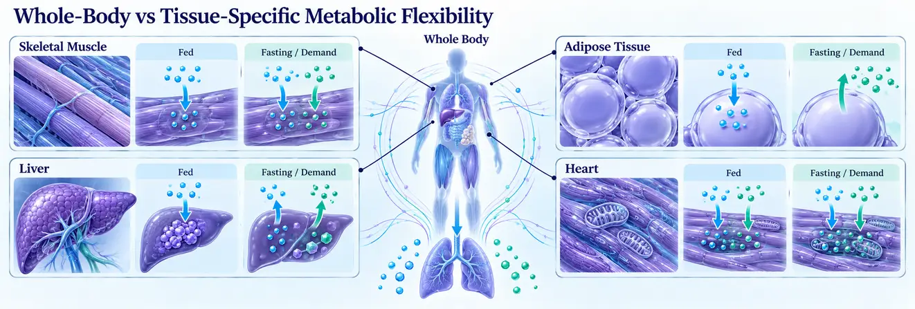
Whole-body gas exchange reflects the combined activity of many organs. Individual tissues may respond differently to the same metabolic challenge, so systemic measurements cannot identify every local change.
Skeletal Muscle
Skeletal muscle is central to insulin-stimulated glucose disposal and can oxidise both carbohydrates and fats. Its response depends on fibre type, mitochondrial capacity, enzyme activity and intracellular stores of glycogen and triglycerides.
The athlete’s paradox illustrates why total muscle lipid content requires careful interpretation. Endurance-trained athletes may have high intramyocellular triglyceride stores alongside high insulin sensitivity, whereas lipid accumulation in insulin-resistant muscle can accompany metabolic impairment. Lipid composition, droplet location and the capacity to mobilise stored fat help explain the difference.
Liver and Adipose Tissue
The liver regulates glycogen storage, glucose production, fatty acid metabolism and ketogenesis. Its response to insulin and nutrient availability affects circulating substrates throughout the body.
Adipose tissue stores and releases fatty acids and produces endocrine signals. Reduced insulin action can disrupt normal suppression of lipolysis, changing the fuel available to muscle and liver. These interactions show why organ-specific dysfunction can affect systemic metabolism.
Heart and Brain
The heart continuously produces ATP from a mixture of fatty acids, glucose, lactate and, under some circumstances, ketones. Its fuel preference changes with workload, substrate availability and disease state; alterations in heart failure are heterogeneous rather than uniform.
The brain generally relies strongly on glucose, although ketone utilisation can rise during prolonged fasting. Red blood cells have a different requirement again, relying on glycolysis because they lack mitochondria.
| Tissue | Major metabolic roles | Research approaches |
| Skeletal muscle | Glucose disposal, glycogen storage, oxidation | Glucose uptake, muscle metabolites, respiration |
| Liver | Glucose production, lipid oxidation, ketogenesis | Isotope tracing, glucose-production studies |
| Adipose tissue | Lipid storage, lipolysis, endocrine signalling | Fatty acid release, adipokine analysis |
| Heart | Continuous oxidation of multiple substrates | Metabolic imaging, tissue respiration |
Explore scientific resources on peptide biology and metabolic research at Australia Peptide Sciences.
Metabolic Flexibility and Insulin Resistance
Metabolic flexibility and insulin resistance are connected within the broader context of metabolic health, but they are not synonymous.
Insulin resistance means a reduced biological response to insulin in one or more tissues. Metabolic inflexibility describes an impaired or inappropriate change in substrate utilisation when conditions change.
How Insulin Resistance Influences Fuel Switching
Insulin normally promotes glucose uptake and shifts metabolism towards carbohydrate utilisation in responsive tissues. With insulin resistance, reduced glucose disposal can limit the increase in carbohydrate oxidation during an insulin challenge.
Researchers often measure this response using indirect calorimetry alongside a hyperinsulinaemic-euglycaemic clamp. A smaller rise in respiratory exchange ratio may indicate a reduced shift towards carbohydrate oxidation. However, it can also result partly from less glucose entering cells, rather than an independent defect in mitochondrial fuel selection.
This distinction matters when comparing people or experimental groups with different insulin sensitivities.
Mitochondrial Dysfunction and Lipid Metabolism
Experimental models associate disrupted insulin signalling with changes in substrate flux, mitochondrial redox balance and lipid-derived metabolites. Some ceramide and diacylglycerol species can interfere with insulin signalling under particular conditions.
Yet increased fatty acid oxidation is not necessarily protective, and reduced oxidation is not always the primary cause of dysfunction. The relationship depends on substrate supply, oxidative capacity and ATP demand. This also helps explain why muscle lipid accumulation may have different implications in trained and insulin-resistant tissues.
What Does Current Research Establish?
A 2025 systematic review and meta-analysis by Hansen and colleagues identified 65 studies using insulin-clamp approaches to assess metabolic flexibility. A subset of 35 comparable studies included 985 participants from lean, overweight or obese, and type 2 diabetes groups.
Average insulin-stimulated changes in respiratory exchange ratio were approximately 0.10 in lean participants and 0.07 in both the overweight or obese and type 2 diabetes groups.
Considerable heterogeneity limited simple comparisons. In the reported meta-regression, body mass index was the participant characteristic significantly associated with the measured response.
The findings challenge the idea of a unique, universal metabolic-inflexibility threshold for type 2 diabetes. They also reinforce the importance of considering glucose disposal and experimental conditions rather than inferring causality from a reduced respiratory response.
How Is Metabolic Flexibility Tested?
A metabolic flexibility test examines changes in fuel utilisation after a controlled nutritional, hormonal or energetic challenge. There is no universal diagnostic test or accepted cut-off applicable to every physiological context.
Indirect Calorimetry and Respiratory Exchange Ratio
Indirect calorimetry measures oxygen consumption (VO₂) and carbon dioxide production (VCO₂). The respiratory exchange ratio is calculated as RER = VCO₂ / VO₂.
Under suitable steady-state conditions, RER approximates the balance of carbohydrate and fat oxidation:
| Approximate RER | Interpretation |
| 0.70 | Predominantly fat oxidation |
| 0.85 | Mixed fat and carbohydrate oxidation |
| 1.00 | Predominantly carbohydrate oxidation |
These are reference values, not diagnostic thresholds. The respiratory quotient (RQ) describes CO₂ production relative to oxygen consumption at the metabolic level; RER is measured through respiratory gas exchange. They approximate one another at steady state but can diverge during rapid physiological changes.
During intense exercise, bicarbonate buffering and changes in ventilation can raise RER above 1.00. A single RER reading therefore cannot establish metabolic flexibility; changes across defined conditions are more informative.
Interpretation also requires attention to protein oxidation and the difference between measured gas exchange and calculated substrate rates. Researchers may use urinary nitrogen measurements or assumptions about protein contribution when estimating non-protein respiratory quotient.
Measurements collected immediately after changing a workload or meal may not represent a steady metabolic state. Calibration, sampling duration, breathing pattern and gas analyser performance can introduce variability. These factors matter particularly when comparing small changes in RER across studies, because an apparent difference may arise from protocol choices rather than altered mitochondrial substrate selection.
Insulin Clamps and Metabolic Challenges
A hyperinsulinaemic-euglycaemic clamp maintains controlled insulin and glucose conditions while investigators measure glucose disposal. Combined with calorimetry, it can quantify changes in substrate oxidation during insulin stimulation.
Fasting-to-fed comparisons, dietary challenges and activity interventions investigate other aspects of fuel switching. Results depend on insulin exposure, nutrient composition, energy balance and participant characteristics. Different challenges may yield different estimates even in the same population.
Whole-Room Calorimetry
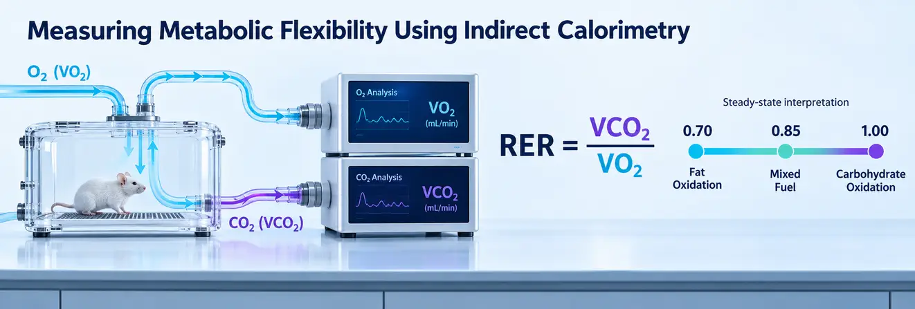
Whole-room indirect calorimetry collects respiratory data over extended periods, allowing researchers to examine substrate oxidation during feeding, sleep and physical activity.
It can reveal patterns obscured by short resting measurements but requires specialised facilities and controlled protocols.
A June 2026 study by Ortega-Santos and colleagues examined metabolic flexibility during controlled dietary and exercise challenges using whole-room calorimetry.
The findings showed that detectability depended on the challenge and index selected. Acute exercise under energy-balanced conditions did not significantly change the indices examined, whereas other nutritional or energy-deficit conditions revealed different responses.
The study illustrates why a larger change in one respiratory metric should not automatically be interpreted as universally better metabolic function.
Cellular Fuel-Flexibility Assays
Extracellular flux analysers measure cellular oxygen consumption rate (OCR) and extracellular acidification-related signals. Specialised assays investigate mitochondrial use of glucose-derived pyruvate, fatty acids and glutamine by restricting selected pathways and observing respiratory compensation.
Three terms should remain distinct:
- Fuel dependency: Baseline reliance on a particular mitochondrial substrate pathway.
- Fuel capacity: Ability to use that pathway when alternatives are inhibited.
- Fuel flexibility: Capacity minus dependency, representing potential additional reliance on that pathway.
Assay interpretation depends on cell type, culture conditions and inhibitor selectivity.
A response in cultured cells cannot be treated as a direct measurement of whole-body metabolic flexibility.
Stable-Isotope Tracing and Metabolomics
Stable-isotope tracing, commonly using carbon-13-labelled substrates, follows labelled atoms into downstream metabolites. Combined with mass spectrometry, nuclear magnetic resonance and suitable computational models, it can help estimate pathway contributions and metabolic flux.
Metabolomics measures patterns of molecules such as organic acids, amino acids, lipids and acylcarnitines. However, metabolite abundance does not necessarily establish pathway flux: concentrations reflect production, consumption and transport. Integrating these methods with respiration measurements provides a more complete experimental picture.
Can Glucose Monitoring Measure Metabolic Flexibility?
Glucose, insulin, ketones, lactate and circulating fatty acids provide useful metabolic context but cannot independently quantify fuel switching. Continuous glucose monitoring measures interstitial glucose dynamics, not mitochondrial substrate oxidation.
Higher ketone concentrations similarly do not prove improved flexibility, because production, utilisation and clearance all affect measured levels.
| Method | Principal measurement | Key limitation |
| Indirect calorimetry | Gas exchange and estimated substrate oxidation | State- and protocol-dependent |
| Insulin clamp plus calorimetry | Glucose disposal and insulin-stimulated fuel switching | Specific hormonal challenge |
| Whole-room calorimetry | Extended respiration and energy expenditure | Specialist infrastructure |
| Cellular fuel assays | Dependency, capacity and flexibility | Cell- and inhibitor-specific |
| Isotope tracing | Labelled substrate movement through pathways | Requires appropriate modelling |
| Glucose and other biomarkers | Selected aspects of metabolic regulation | Not direct measures of overall flexibility |
Metabolic Flexibility Diet: What Does Research Show?
A metabolic flexibility diet is not a single scientifically standardised nutritional intervention. Researchers instead test how macronutrient composition, nutrient timing and energy availability affect fuel utilisation.
Carbohydrates, Fat and Substrate Adaptation
Greater carbohydrate availability generally favours carbohydrate oxidation; higher dietary fat availability can increase fat oxidation. Neither response automatically demonstrates improved flexibility. A change may simply reflect which substrate is more abundant.
Results differ across high-fat and high-carbohydrate feeding studies according to intervention duration, energy balance, insulin sensitivity and the outcome measured.
Substrate preference under one diet must be distinguished from the capacity to adjust when conditions change.
Fasting and Energy Restriction
Fasting can increase fatty acid mobilisation and ketone production, while nutrient scarcity influences cellular regulators including AMPK. Nevertheless, increased fat oxidation during fasting does not establish a lasting improvement in metabolic adaptability.
A 2025 randomised trial by Oldenburg and colleagues compared time-restricted eating, calorie restriction and unrestricted eating over 12 weeks.
It did not demonstrate significant overall advantages for time-restricted eating in the principal metabolic outcomes, and clamp-measured flexibility was lower than in the calorie-restriction group.
The result should not be generalised to every fasting protocol. Findings from preclinical time-restricted feeding studies also require separate interpretation, as animal and human studies do not necessarily produce equivalent outcomes. Changes in diet, glucose regulation and measured metabolic flexibility cannot be assumed to follow the same pattern.
Why Findings Differ
Dietary studies vary in duration, energy balance, macronutrient composition, baseline metabolic status and testing method. Animal and cell models introduce additional limits to translation. A fasting RER, insulin-stimulated RER and 24-hour RQ profile each describes a different aspect of fuel utilisation, so they should not be treated as interchangeable endpoints.
Can Metabolic Flexibility Be Improved? Research Evidence
Research on how to improve metabolic flexibility examines changes in mitochondrial capacity, insulin responsiveness and measured substrate switching. Demonstrating improvement in one component is not proof that all aspects of metabolism have improved.
Exercise-Related Adaptation
Endurance training can increase skeletal muscle oxidative capacity and alter enzymes involved in glucose and fatty acid metabolism. Muscle contraction also stimulates glucose transport partly independently of insulin; longer-term training can affect GLUT4 expression and glycogen handling.
These adaptations may change responses to metabolic challenges, but an acute exercise response differs from a training adaptation. The 2026 calorimetry findings, for instance, did not show significant changes in the selected flexibility indices after acute exercise under energy-balanced conditions.
Energy Balance and Peptide Signalling Research
Energy restriction and changes in body composition can alter insulin sensitivity and substrate oxidation, but separating these effects from changes in glucose disposal or mitochondrial function is difficult. Greater fat oxidation alone does not confirm improved switching towards glucose when required.
Peptide hormones are studied in experimental models to investigate metabolic regulation across tissues.
GLP-1 and GIP contribute to nutrient-responsive signalling, including pathways investigated using dual-receptor agonists such as Tirzepatide. Glucagon influences hepatic glucose production and ketogenesis, while fibroblast growth factor 21 (FGF21) participates in metabolic stress responses.
Genetic and pharmacological studies can help identify mechanisms, but changes in glucose or circulating metabolites do not alone demonstrate improved whole-body flexibility.
Results involving experimental compounds must remain distinguished from established clinical outcomes.
Current Limitations and Future Directions
Standardising Measurements
Studies define metabolic flexibility through insulin-stimulated RER changes, dietary responses, exercise-associated oxidation or extended respiratory patterns. Each tests a different challenge. Nutrition, activity, adiposity, energy balance and measurement duration influence results, limiting comparisons.
A 2026 review of whole-room calorimetry emphasised the importance of consistent protocols and interpretation. Greater standardisation would help determine whether differences reflect biological capacity or measurement design.
Emerging Research Methods

Isotope-based flux analysis and high-resolution metabolomics can identify pathway changes not evident from blood concentrations alone.
Positron emission tomography uses selected radiotracers to investigate tissue-specific substrate uptake. Hyperpolarised carbon-13 magnetic resonance methods can examine selected transformations, including pyruvate metabolism.
These techniques offer complementary mechanistic information but are not validated stand-alone measures of complete metabolic flexibility. Integrating them with physiological challenges may better connect cellular changes to whole-body responses.
Explore our research peptide shop for compounds intended exclusively for laboratory research.
Unresolved Questions
Researchers still need to determine whether inflexibility precedes insulin resistance or develops alongside it, which assays independently predict meaningful outcomes, and how closely cultured-cell results reflect intact organisms. Establishing these relationships requires controlled studies and clear distinctions between association and causation.
Understanding Metabolic Flexibility: Key Research Findings
Metabolic flexibility is the capacity to adapt fuel utilisation to changing nutrient availability and energy needs. It depends on mitochondrial pathways, hormonal signals and communication between metabolically specialised tissues.
Although impaired flexibility often accompanies insulin resistance, the two are not interchangeable and their causal relationship remains unresolved. Indirect calorimetry, controlled challenges and cellular analyses each provide useful but incomplete evidence.
Reliable conclusions require interpreting the measurement alongside the biological model and experimental conditions.
FAQs About Metabolic Flexibility
How Is Metabolic Flexibility Assessed in Research?
Metabolic flexibility cannot be determined reliably from everyday symptoms or energy levels. Researchers assess it by measuring changes in substrate oxidation during controlled metabolic challenges, such as fasting, feeding or insulin stimulation. There is currently no universally accepted diagnostic threshold.
How Can Metabolic Flexibility Be Measured?
Metabolic flexibility is commonly investigated using indirect calorimetry, which measures oxygen consumption and carbon dioxide production to estimate fuel oxidation.
Changes in respiratory exchange ratio (RER) across controlled metabolic states reveal how substrate utilisation responds. Other research methods include insulin clamp studies, whole-room calorimetry and cellular respiration assays.
What Are the Signs of Metabolic Inflexibility?
Metabolic inflexibility involves a reduced or inappropriate change in fuel utilisation when metabolic conditions change. In laboratory studies, it may appear as a smaller shift towards carbohydrate oxidation during insulin stimulation. Although associated with insulin resistance and altered mitochondrial metabolism, it cannot be reliably identified through symptoms alone.
How Can Metabolic Flexibility Be Improved?
Research indicates that metabolic flexibility can change through adaptations in mitochondrial function, insulin signalling and substrate metabolism.
Controlled exercise and nutritional studies have demonstrated such changes, although results vary according to the experimental conditions. Increased fat oxidation alone does not establish improved metabolic flexibility.
What Is the Relationship Between Metabolic Flexibility and Insulin Resistance?
Insulin resistance can reduce glucose uptake and limit the normal increase in carbohydrate oxidation during insulin stimulation.
However, metabolic flexibility also depends on mitochondrial activity, hormonal regulation and tissue-specific metabolism. The two are associated, but their causal relationship remains incompletely understood.
Can Exercise Improve Metabolic Flexibility and Insulin Sensitivity?
Exercise training can increase skeletal muscle oxidative capacity, improve glucose transport and enhance insulin responsiveness.
These adaptations may influence how effectively tissues adjust substrate utilisation. However, improvements in insulin sensitivity do not always correspond to measurable increases in metabolic flexibility.
Is Aerobic or Resistance Exercise More Effective for Metabolic Flexibility?
Aerobic training has been extensively investigated for its effects on mitochondrial oxidative capacity and fatty acid metabolism.
Resistance training also influences skeletal muscle glucose uptake and insulin sensitivity. Current evidence does not establish either approach as universally superior for metabolic flexibility, partly because studies employ different measurement methods.
Does Intermittent Fasting Improve Metabolic Flexibility?
Intermittent fasting alters nutrient availability and generally increases reliance on fatty acid oxidation during fasting periods. However, this temporary change does not necessarily indicate improved fuel-switching capacity. Controlled human studies have reported mixed findings, suggesting that outcomes depend on the intervention and assessment method.
How Does Diet Affect Metabolic Flexibility?
Dietary composition influences the availability and oxidation of carbohydrates and fats. Greater carbohydrate availability generally promotes glucose oxidation, while increased fat availability can shift substrate utilisation towards fatty acids. Whether these changes reflect improved metabolic flexibility depends on energy balance, metabolic status and the capacity to adapt to subsequent nutritional challenges.
Can Metabolic Flexibility Be Improved Without a Ketogenic Diet?
Yes. Ketogenic feeding is not a necessary condition for metabolic flexibility, which describes the ability to adapt fuel utilisation rather than maintain a particular metabolic state.
Research investigates this capacity through various dietary challenges, exercise-related adaptations and changes in energy availability without requiring sustained ketosis.
What Role Do Mitochondria Play in Metabolic Flexibility?
Mitochondria support ATP production by oxidising substrates derived from carbohydrates, fatty acids and other nutrients.
Their transport systems and metabolic enzymes help adjust fuel utilisation according to cellular energy requirements. Mitochondrial function is central to metabolic flexibility, although hormonal signals and substrate availability also influence the response.
Does Better Fat Burning Mean Greater Metabolic Flexibility?
No. Increased fatty acid oxidation indicates greater fat utilisation under particular conditions, not necessarily improved metabolic flexibility.
A flexible metabolic system must also adjust appropriately when carbohydrate availability or energy demand changes. The capacity to switch between substrates is more informative than the oxidation rate of one fuel alone.
Can Blood Glucose Levels Indicate Metabolic Flexibility?
Blood glucose measurements reveal aspects of glucose regulation but do not directly measure carbohydrate or fatty acid oxidation.
Continuous glucose monitoring can track changes in interstitial glucose concentrations, whereas metabolic flexibility requires assessing substrate utilisation under different conditions. Glucose measurements may complement research testing but cannot independently establish metabolic flexibility.