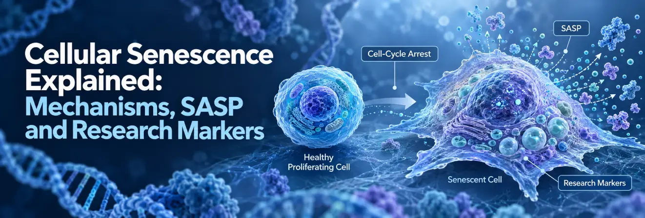
Cellular senescence is a biological state in which cells enter stable cell-cycle arrest, commonly following DNA damage, telomere dysfunction or other cellular stress. Unlike cells undergoing apoptosis, senescent cells generally remain viable and metabolically active, although their structure, gene expression and interactions with neighbouring cells change.
Senescence can suppress the proliferation of potentially damaged cells and contribute to tissue remodelling and repair. Yet persistent senescent cell populations may alter inflammatory signalling and tissue function. Their effects depend on cell type, molecular pathways and environmental context.
Researchers investigate these responses using cell-cycle regulators, biochemical markers, secreted molecules and experimental models. Understanding their limitations is essential because senescent cells do not share one universal molecular signature.
What Is Cellular Senescence?s
Cellular senescence is defined by sustained proliferative arrest accompanied by molecular and functional changes that distinguish senescent cells from other non-dividing cells. Early experiments showed that normal human fibroblasts have a finite capacity to divide in culture. Later research demonstrated that senescence can also arise without repeated division, including through oxidative stress, oncogenic signalling and persistent DNA damage.
What Defines a Senescent Cell?
Senescent cells commonly exhibit:
- Stable cell-cycle arrest: Resistance to ordinary physiological signals that promote cell division.
- Continued viability: Ongoing metabolism despite the loss of proliferative capacity.
- Morphological changes: Enlarged or flattened shapes, particularly in cell culture.
- Molecular remodelling: Changes in stress-response genes, chromatin and cell-cycle regulators.
- Altered secretion: In many cells, production of a senescence-associated secretory phenotype (SASP).
None of these accompanying features is present in every senescent population. Phenotypes vary with the initiating stress, tissue and stage of senescence. Researchers also observe senescence-like changes in normally non-dividing neurons and cardiomyocytes, although classical proliferative arrest cannot be assessed in those cells in the same way.
Cellular Senescence vs Quiescence, Apoptosis and Differentiation
Senescence differs from other states associated with reduced cell division.
| Cellular state | Main characteristic | Key distinction |
| Senescence | Stable arrest with molecular and functional changes | Cells generally resist normal growth signals |
| Quiescence | Temporary withdrawal from proliferation | Cells can typically resume division |
| Apoptosis | Regulated cell death | The cell is eliminated |
| Terminal differentiation | Acquisition of specialised functions | Reduced proliferation is linked to maturation |
A lack of proliferation markers alone cannot establish senescence: quiescent and differentiated cells may show similar results. Researchers therefore assess several independent features.
One further distinction concerns reversibility. Quiescent cells usually retain an ordinary capacity to resume division, while senescent cells show arrest that is difficult to overcome through physiological stimulation. Nevertheless, the molecular depth of senescence can change over time, and loss of checkpoint control may allow exceptional cells to escape arrest. This does not make the states interchangeable.
Discover Australia Peptide Sciences and explore our research resources.
What Causes Cellular Senescence?
Different biological stresses can initiate senescence, often through overlapping cell-cycle checkpoint pathways.
Replicative Senescence and Telomere Shortening
Replicative cell senescence describes the loss of proliferative capacity in many normal somatic cells after successive divisions. It is closely associated with telomeres, protective DNA-protein structures at chromosome ends.
In many somatic cells, telomeres shorten because conventional DNA replication cannot fully reproduce chromosome ends. Critically short or structurally unprotected telomeres can activate a persistent DNA damage response (DDR), leading to checkpoint signalling and cell-cycle arrest.
The historical Hayflick limit describes the finite division capacity of cultured normal cells. Although around 50 divisions is frequently cited for certain human fibroblasts, the number varies with cell type and experimental conditions.
Telomerase can help maintain telomeric DNA in some cell populations, but its presence does not prevent every stress-induced senescence response. Telomere dysfunction can also occur without substantial shortening, so length alone cannot determine senescence status.
In telomere research, colocalisation of telomeric signals with DNA damage proteins can provide evidence of telomere-associated DNA damage beyond telomere length measurements alone. Separately, cell-culture studies involving Epitalon have investigated telomerase-related endpoints, although these findings do not independently establish cellular senescence.
DNA Damage, Oxidative Stress and Organelle Dysfunction
Cells continually monitor genetic integrity through DDR pathways. When damage occurs, these pathways coordinate repair and temporarily restrict proliferation. Persistent or unresolved lesions may instead contribute to sustained arrest.
Reactive oxygen species (ROS) participate in normal signalling, but excessive production or impaired redox regulation can damage DNA, proteins and lipids. Experimental models connect oxidative stress with senescence-associated signalling through p38 MAPK, p53 and related regulators.
Mitochondrial dysfunction can alter energy metabolism and redox balance, both relevant to NAD+ research. Lysosomal abnormalities and disrupted protein homeostasis can also reinforce cellular stress. Their contributions vary across cell types and models rather than forming one obligatory sequence
Researchers may assess glutathione -related redox status alongside ROS measurements to investigate whether oxidative changes precede or follow cell-cycle arrest. A correlation between high ROS and senescence markers, for example, does not establish that oxidative stress initiated the response. Perturbation experiments and longitudinal measurements are needed to investigate the direction of that relationship.
Oncogene-Induced and Other Forms of Senescence
Oncogene-induced senescence (OIS) can arise when abnormal growth-promoting signals provoke replication stress and checkpoint activation. Excessive RAS signalling is a well-studied example. This response may restrict proliferation in cells with potentially oncogenic alterations, although it depends on intact regulatory pathways.
Senescence-like programmes are also observed after certain genotoxic exposures and during developmental tissue remodelling.
| Senescence type | Main trigger | Molecular response | Typical research model |
| Replicative | Telomere dysfunction after repeated division | Persistent DDR and arrest | Primary cell cultures |
| DNA damage-induced | Unresolved genomic lesions | Checkpoint activation | Genotoxic stress models |
| Oxidative stress-induced | Altered redox balance | Stress signalling and arrest | Cellular stress experiments |
| Oncogene-induced | Abnormal mitogenic signalling | Replication stress and tumour-suppressor activation | Oncogene activation models |
| Developmental | Regulated developmental signals | Tissue-specific programmes | Developmental models |
These categories overlap because different triggers can engage shared pathways.
How Does Cellular Senescence Work at the Molecular Level?
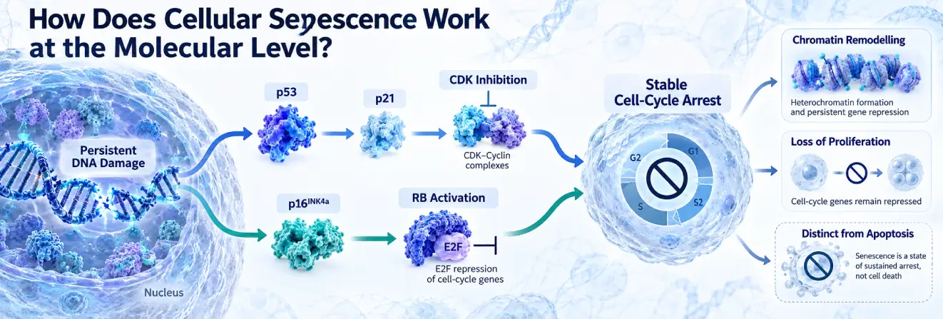
The p53-p21 and p16INK4a-RB pathways are central regulators of senescence-associated cell-cycle arrest, although neither operates identically in every cell population.
The p53-p21 Pathway in Senescence
The relationship between p53 and senescence helps explain how persistent stress restricts proliferation. p53, encoded by TP53, regulates genes involved in cell-cycle control, DNA repair and apoptosis.
Following DNA damage, signalling networks involving ATM and ATR help regulate p53. Activated p53 can increase transcription of CDKN1A, which encodes the cyclin-dependent kinase inhibitor p21. p21 inhibits cyclin-dependent kinases (CDKs), limiting progression through key cell-cycle checkpoints.
Persistent DNA damage → DDR signalling → p53 activation → p21 expression → CDK inhibition → cell-cycle arrest.
This sequence is a simplified model, not a universal outcome. Depending on the stress and cellular context, p53 can promote cell-cycle arrest or apoptosis. It can also contribute to senescence initiation without remaining equally important for its maintenance.
The p16INK4a-RB Pathway
p16INK4a, encoded by CDKN2A, inhibits CDK4 and CDK6. These kinases ordinarily phosphorylate retinoblastoma protein (RB), permitting E2F-dependent transcription of genes needed for proliferation.
By limiting CDK4/6 activity, p16 helps maintain RB in a growth-suppressive state and restricts cell-cycle progression. In some senescent populations, this pathway reinforces arrest initiated by other stress responses.
The CDKN2A locus also produces ARF through an alternative transcript. ARF can influence p53 stability through MDM2, linking the regulatory networks. However, expression of p16 and p21 differs across models; neither is universally elevated in every senescent cell.
Because RB and p53 control more than senescence, their activation must be evaluated in context. For instance, transient p21 elevation may reflect a repair-associated pause rather than permanent proliferative arrest. Repeated measurements and additional evidence of senescence-associated remodelling make mechanistic conclusions more convincing.
Chromatin Remodelling and Maintenance of Senescence
Senescence may involve extensive changes in chromatin structure and transcription. Some models develop senescence-associated heterochromatin foci (SAHF), condensed regions linked to altered expression of proliferation-related genes.
Other changes include histone modifications, redistribution of chromatin-associated proteins and reduced lamin B1, a component of the nuclear lamina. These changes may help stabilise arrest, but SAHF formation and lamin B1 reduction are not universal senescence features.
What Changes Occur in Senescent Cells?
Senescence can affect cell shape, organelle activity, metabolism and nuclear organisation, as well as proliferation.
Morphological and Metabolic Changes
Cultured senescent cells often appear enlarged, flattened and more granular than dividing cells. Some models also show increased mitochondrial mass, altered respiration, elevated ROS or changes in glycolysis and lipid metabolism.
Changes in mTOR, AMPK and autophagy-related pathways have been reported, but their direction and importance vary with cell type and trigger. Increased mitochondrial abundance does not necessarily mean improved mitochondrial function.
Nuclear and Intracellular Changes
Some senescent cells display reduced lamin B1, chromatin reorganisation and persistent DDR signalling. Nuclear γH2AX foci can indicate DNA damage signalling, while increased lysosomal content may accompany changes in intracellular turnover.
Altered apoptosis resistance has also been observed in certain senescent populations. These traits are informative but not exclusive: other stressed or non-dividing cells can display similar changes.
Importantly, senescent-cell morphology is easier to recognise in two-dimensional culture than in intact tissues, where neighbouring structures constrain cell shape. Laboratory interpretation therefore benefits from comparing senescent candidates with appropriate controls from the same cell type and tissue environment.
What Is the Senescence-Associated Secretory Phenotype (SASP)?
The senescence-associated secretory phenotype (SASP) describes changes in the molecules released by many senescent cells. These factors influence immune responses, tissue organisation and neighbouring-cell behaviour beyond the arrested cell itself.
SASP Components and Their Biological Functions
Researchers investigate several classes of secreted factors:
- Cytokines, including IL-6 and IL-1 family members, involved in inflammatory signalling.
- Chemokines, including CXCL8, which influence immune-cell recruitment.
- Growth factors that affect cell communication and tissue responses.
- Matrix metalloproteinases (MMPs) involved in extracellular matrix remodelling.
- Other mediators, including extracellular vesicles and bioactive lipids.
These examples are not a universal SASP signature. The composition, amount and biological effect of secretion depend on the cell population and its environment.
How SASP Influences Neighbouring Cells
Through paracrine signalling, senescent cells can influence nearby immune, stromal and progenitor cells. In some settings, SASP factors attract immune cells that participate in senescent-cell clearance or support tissue remodelling during repair.
Persistent secretory activity may instead sustain inflammatory responses or induce senescence-associated changes in neighbouring cells. Whether the outcome is protective or disruptive depends on the secreted factors, tissue and duration of signalling.
Why SASP Varies Between Senescent Cell Populations
Cell identity, inducing stress, tissue environment and time since senescence onset all shape SASP composition. NF-κB and other stress-responsive pathways regulate aspects of inflammatory secretion in several models.
Not every senescent cell develops a strongly inflammatory SASP. Conversely, detecting one cytokine cannot establish cellular senescence because the same molecule may be released during other cellular responses.
Secreted proteins can also act differently depending on their local concentrations and the responsiveness of neighbouring cells. Researchers increasingly examine secretory profiles as coordinated molecular patterns rather than interpreting individual cytokines as universal indicators of inflammation or senescence.
How Do Researchers Identify Cellular Senescence?
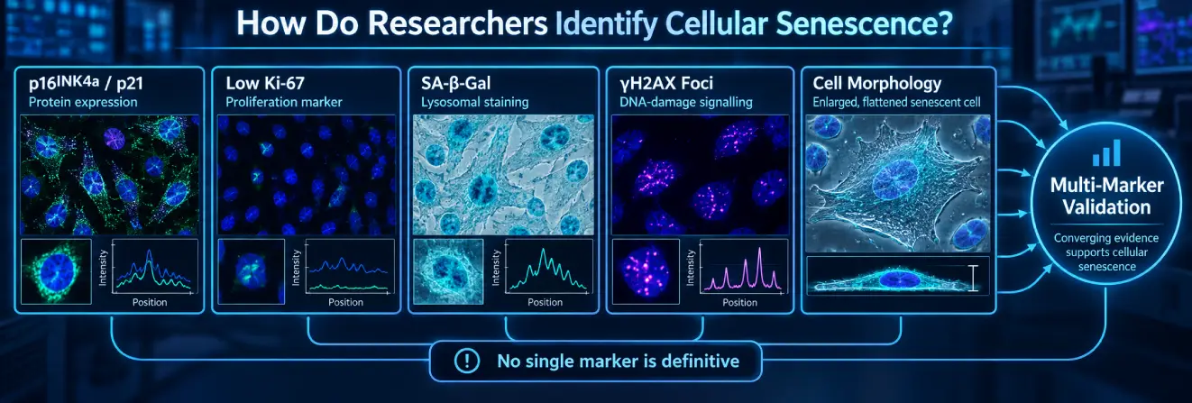
No individual laboratory marker reliably identifies every senescent cell. Researchers combine evidence of cell-cycle regulation with morphological, biochemical and molecular observations, accounting for differences between cell cultures and intact tissues.
Cell-Cycle Arrest and Molecular Markers
p16INK4a and p21 are widely studied cell-cycle inhibitors. Their expression can be examined through immunostaining, immunoblotting and RNA analysis, but neither alone confirms senescence.
Researchers also assess proliferation using Ki-67 staining or nucleotide-incorporation assays such as EdU and BrdU. Low proliferation supports evidence of arrest but cannot distinguish senescence from quiescence or differentiation. Where feasible, responses to renewed growth signals add useful functional information.
A time-course design can help distinguish sustained arrest from a short-lived response following stress. In mixed populations, investigators must also consider whether strongly proliferating cells obscure smaller senescent subpopulations. Measurements at the single-cell level can address this limitation more effectively than averages across a whole culture.
SA-β-Gal Staining and Its Limitations
Senescence-associated β-galactosidase (SA-β-gal) is a common laboratory marker. The assay measures elevated lysosomal β-galactosidase activity under conditions typically adjusted to approximately pH 6.0. This activity is associated with the enzyme encoded by GLB1.
Histochemical methods produce visible staining, while suitable fluorescence-based methods provide alternative readouts. Many senescent cells show increased activity because of altered or expanded lysosomal compartments.
However, high SA-β-gal activity is not unique to senescence. Certain non-senescent, quiescent or highly lysosomal cells may also test positive, and culture conditions can influence results. SA-β-gal staining should therefore be interpreted alongside independent markers.
Additional Biomarkers and Multi-Marker Validation
Different markers measure different aspects of the senescent phenotype.
| Marker | Biological feature | Common method | Main limitation |
| SA-β-gal | Lysosomal enzyme activity | Histochemical or fluorescence assay | Not senescence-specific |
| p16INK4a | Cell-cycle inhibition | Immunostaining or RNA analysis | Cell-type-dependent expression |
| p21 | Stress-responsive arrest | Immunostaining or RNA analysis | May increase during temporary stress |
| Ki-67 | Proliferation | Immunostaining | Low expression is not diagnostic |
| γH2AX | DNA damage signalling | Immunofluorescence | Also appears outside senescence |
| Lamin B1 | Nuclear lamina organisation | Immunostaining or immunoblotting | Reduction is not universal |
| SASP factors | Secretory activity | ELISA, proteomics or transcriptomics | Profiles vary by context |
The 2024 MICSE guidelines emphasise combining markers. In relevant tissue-based settings, evaluating p21 and/or p16 together with at least two additional indicators from different senescence-associated categories can strengthen interpretation.
Co-detection within individual cells is particularly valuable. For example, elevated p21 alongside reduced lamin B1 and increased SA-β-gal offers stronger support than any result alone. Nevertheless, identifying senescence-associated features is different from proving that those cells cause a specific tissue outcome; causal claims require functional experiments.
Marker combinations should also reflect the model under investigation. A marker panel validated in cultured fibroblasts may perform differently in immune cells or aged tissue. Appropriate positive and negative controls, assay-specific background measurements and transparent reporting help researchers distinguish genuine cellular changes from technical effects.
Watch this video to understand how senescent cells form and how researchers identify them using molecular markers.
What Role Does Cellular Senescence Play in Ageing, Tissue Repair and Cancer?
Senescence can have protective or disruptive consequences depending on its timing, persistence and biological setting.
Protective Roles in Development and Tissue Repair
Stable arrest can prevent proliferation of cells containing potentially oncogenic damage, providing an important tumour-suppressive mechanism.
Transient senescence-associated programmes also participate in some developmental and tissue-remodelling processes. In experimental repair models, secreted factors can influence immune recruitment and extracellular matrix organisation. Subsequent immune-mediated clearance may help limit the persistence of these cells.
Persistent Senescence and Age-Related Changes
Some senescent populations become more prevalent with age. Possible contributors include cumulative cellular stress, repeated senescence induction and changes in immune clearance.
Animal experiments have associated selected persistent populations with inflammation, altered metabolism and impaired tissue maintenance. However, these findings do not mean every senescent cell is harmful or that senescence alone causes biological ageing.
Cellular ageing also involves genomic instability, changes in nutrient sensing, mitochondrial function and intercellular communication.
Human associations should be distinguished from causal effects demonstrated in experimental models.
Transient and persistent senescence are not defined simply by the age of the organism. A temporary response may develop later in life, while unresolved senescence-associated signals may arise in younger tissues under particular pathological or experimental conditions. Duration, tissue location and immune surveillance remain central variables.
The Dual Role of Senescence in Cancer Biology
Senescence can suppress tumour development by arresting potentially abnormal cells. Within established tumour environments, however, senescent stromal or other cell populations may release factors that influence inflammation, immune behaviour and extracellular matrix organisation.
Some experimental models link these changes to conditions that support tumour progression. The outcome depends on cell type, secretory profile and local tissue context; senescence is not uniformly tumour-suppressive or tumour-promoting.
How Is Cellular Senescence Studied in Laboratory Research?
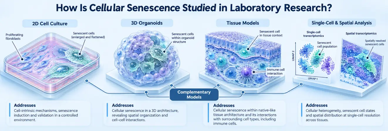
Researchers use complementary model systems to distinguish the initiation of senescence, maintenance of arrest and interactions with surrounding cells.
Cell-Culture and Experimental Models
Primary fibroblast cultures remain useful for studying replicative senescence, including telomere-related changes. Other models examine responses to DNA damage, oncogenic activation or organelle stress. In studies investigating specific molecular pathways, cellular research peptides may also be examined under controlled laboratory conditions, although their relevance to senescence must be established through experimental evidence.
Cell cultures allow detailed analysis of proteins, gene expression and metabolism but do not reproduce the complete cellular environment of intact tissues. Three-dimensional cultures and organoids provide additional context, while animal models allow investigation of senescence-associated processes across interacting cell populations. Species differences and model-specific effects still limit extrapolation.
Different models also capture different stages of senescence. An acute laboratory stress response may produce a phenotype over a shorter period than the gradual changes observed in tissues. Reporting the timing of measurements, cellular background and definition of senescence makes results more reproducible and avoids assuming that distinct models are equivalent.
Single-Cell and Spatial Research Techniques
Single-cell and single-nucleus multi-omics approaches can reveal molecular differences between cell populations, helping researchers characterise senescence-associated states. Spatial transcriptomics adds information about where those populations are located and how they relate to neighbouring cells.
Proteomics and other multi-omics approaches provide complementary information about signalling and metabolic changes. Yet computational senescence signatures may overlap with inflammation or differentiation; molecular classifications require independent experimental validation.
Spatial information is particularly helpful when a biological effect might result from interactions between senescent cells and neighbouring immune or stromal populations. Combining cellular location with protein detection and functional measurements can provide stronger evidence than transcriptomic enrichment alone.
Explore research peptides at Australia Peptide Sciences.
Emerging Research Directions and Unanswered Questions
Current research increasingly focuses on the heterogeneity of senescent cells and the biological consequences of their persistence.
Experimental Senolytic and Senomorphic Research
Senolytic research examines approaches intended to preferentially eliminate senescent cells through vulnerabilities in their survival mechanisms. Senomorphic research instead investigates modifications to senescence-associated activity, including secretory signalling, without necessarily removing the cells.
Preclinical effects differ between cell populations and models. Reduced expression of one marker does not prove cell clearance or reversal of senescence. Early clinical investigations have not established broad clinical efficacy or long-term safety for senescence-targeting approaches.
Current Challenges in Senescence Research
The absence of universal markers complicates comparisons between studies. Researchers also investigate whether different senescent states remain stable, how immune clearance changes over time and which populations support repair versus persistent tissue dysfunction.
Some experimental cells can escape arrest when checkpoint pathways are disrupted, but changes in SASP or marker expression alone are not evidence of restored proliferation. Better longitudinal models, cell-specific markers and functional validation are needed to resolve these questions.
Another important question is how immune surveillance selects senescent targets. Surface proteins, secreted signals and tissue-specific immune conditions may all contribute, but findings from one experimental setting cannot automatically be generalised to others. Characterising these interactions could help researchers distinguish normal clearance processes from persistent accumulation.
Understanding the Biology of Cellular Senescence
Cellular senescence combines stable proliferative arrest with context-dependent changes in metabolism, chromatin and cell signalling. Telomere dysfunction, DNA damage, oxidative stress and oncogenic signalling can activate pathways involving p53, p21, p16INK4a and RB.
Senescent cells may support tumour suppression and tissue repair, while persistent populations can contribute to altered inflammatory responses. Their heterogeneity makes multi-marker laboratory assessment essential. Emerging molecular and spatial techniques are improving how researchers distinguish senescent cell populations and investigate their functional effects.
FAQs About Cellular Senescence
What Is Cellular Senescence?
Cellular senescence occurs when a cell stops dividing in response to biological stress but continues performing essential metabolic functions.
This state is commonly associated with DNA damage, telomere dysfunction and changes in cell-cycle regulation. Senescent cells may also develop distinctive molecular characteristics, including altered gene expression and secretion of signalling molecules that influence neighbouring cells.
Is Cellular Senescence Good or Bad?
The effects of cellular senescence depend on when and where it occurs. By restricting the proliferation of damaged cells, senescence contributes to tumour suppression and certain tissue-repair processes. However, when senescent cells persist, their altered signalling and inflammatory secretions may disrupt tissue function. These contrasting effects make cellular senescence an important subject in ageing and molecular biology research.
How Are Senescent Cells Cleared?
Senescent cells can be removed through natural immune surveillance, involving immune cells such as macrophages, natural killer cells and cytotoxic T cells. In laboratory research, scientists investigate senolytic compounds designed to preferentially eliminate senescent cells. Other experimental approaches examine ways to modify their secretory activity. The effectiveness and selectivity of these strategies depend on the biological model, and their clinical benefits remain uncertain.
What Triggers Cell Senescence?
Cellular senescence can develop when persistent stress interferes with normal cellular regulation. Common triggers include progressive telomere shortening, unresolved DNA damage, excessive reactive oxygen species and abnormal oncogene activation. These conditions can engage signalling pathways involving p53, p21 and p16INK4a, leading to sustained cell-cycle arrest. Different cell populations may respond to the same stressor through distinct molecular mechanisms.
At What Age Does Senescence Start?
There is no specific age at which cellular senescence begins. Senescence-associated processes can occur during early development and continue throughout life in response to physiological signals or cellular damage. Although senescent cell populations tend to increase in certain tissues with advancing age, their prevalence depends on factors such as cellular stress, tissue characteristics and immune-mediated clearance.
What Speeds Up Cellular Aging?
Cellular ageing can be influenced by accumulated genetic damage, oxidative stress, mitochondrial dysfunction and declining mechanisms of cellular maintenance. Persistent exposure to these stressors may disrupt protein homeostasis, DNA repair and metabolic regulation, increasing the likelihood of senescence in susceptible cells. However, cellular ageing involves multiple biological processes, and not every age-associated cellular change results in senescence.
{
“@context”: “https://schema.org”,
“@type”: “FAQPage”,
“mainEntity”: [
{
“@type”: “Question”,
“name”: “What Is Cellular Senescence?”,
“acceptedAnswer”: {
“@type”: “Answer”,
“text”: “Cellular senescence occurs when a cell stops dividing in response to biological stress but continues performing essential metabolic functions. This state is commonly associated with DNA damage, telomere dysfunction and changes in cell-cycle regulation. Senescent cells may also develop distinctive molecular characteristics, including altered gene expression and secretion of signalling molecules that influence neighbouring cells.”
}
},
{
“@type”: “Question”,
“name”: “Is Cellular Senescence Good or Bad?”,
“acceptedAnswer”: {
“@type”: “Answer”,
“text”: “The effects of cellular senescence depend on when and where it occurs. By restricting the proliferation of damaged cells, senescence contributes to tumour suppression and certain tissue-repair processes. However, when senescent cells persist, their altered signalling and inflammatory secretions may disrupt tissue function. These contrasting effects make cellular senescence an important subject in ageing and molecular biology research.”
}
},
{
“@type”: “Question”,
“name”: “How Are Senescent Cells Cleared?”,
“acceptedAnswer”: {
“@type”: “Answer”,
“text”: “Senescent cells can be removed through natural immune surveillance, involving immune cells such as macrophages, natural killer cells and cytotoxic T cells. In laboratory research, scientists investigate senolytic compounds designed to preferentially eliminate senescent cells. Other experimental approaches examine ways to modify their secretory activity. The effectiveness and selectivity of these strategies depend on the biological model, and their clinical benefits remain uncertain.”
}
},
{
“@type”: “Question”,
“name”: “What Triggers Cell Senescence?”,
“acceptedAnswer”: {
“@type”: “Answer”,
“text”: “Cellular senescence can develop when persistent stress interferes with normal cellular regulation. Common triggers include progressive telomere shortening, unresolved DNA damage, excessive reactive oxygen species and abnormal oncogene activation. These conditions can engage signalling pathways involving p53, p21 and p16INK4a, leading to sustained cell-cycle arrest. Different cell populations may respond to the same stressor through distinct molecular mechanisms.”
}
},
{
“@type”: “Question”,
“name”: “At What Age Does Senescence Start?”,
“acceptedAnswer”: {
“@type”: “Answer”,
“text”: “There is no specific age at which cellular senescence begins. Senescence-associated processes can occur during early development and continue throughout life in response to physiological signals or cellular damage. Although senescent cell populations tend to increase in certain tissues with advancing age, their prevalence depends on factors such as cellular stress, tissue characteristics and immune-mediated clearance.”
}
},
{
“@type”: “Question”,
“name”: “What Speeds Up Cellular Aging?”,
“acceptedAnswer”: {
“@type”: “Answer”,
“text”: “Cellular ageing can be influenced by accumulated genetic damage, oxidative stress, mitochondrial dysfunction and declining mechanisms of cellular maintenance. Persistent exposure to these stressors may disrupt protein homeostasis, DNA repair and metabolic regulation, increasing the likelihood of senescence in susceptible cells. However, cellular ageing involves multiple biological processes, and not every age-associated cellular change results in senescence.”
}
}
]
}