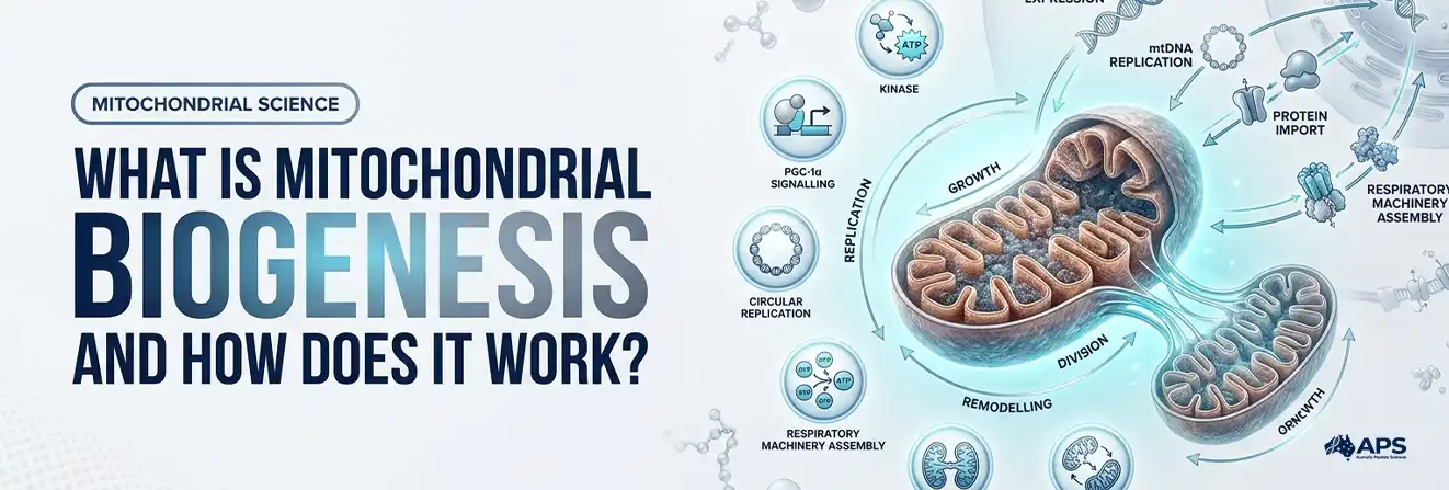
Mitochondrial biogenesis is the coordinated process through which cells expand and renew their mitochondrial network.
The process requires more than mitochondrial division. Cells must activate nuclear genes, replicate mitochondrial DNA (mtDNA), produce proteins and membrane lipids, import nuclear-encoded proteins, and assemble functional respiratory machinery. This may increase mitochondrial content, improve the cell’s capacity to meet energy demands, or achieve both outcomes.
It occurs at a baseline rate and can increase during development and tissue repair, or in response to repeated muscle contraction, cold exposure, and changes in energy availability. The scale of the response depends on the tissue, training status, age, and experimental conditions.
What Does Biogenesis Mean in Mitochondrial Biology?
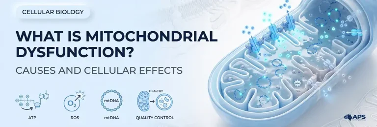
Mitochondria are dynamic, double-membrane organelles that produce much of the adenosine triphosphate (ATP) used by cells through oxidative phosphorylation. They also regulate calcium balance, redox signalling, metabolism, and programmed cell death. Together, these processes are central to mitochondrial function and cellular health.
Biogenesis describes the expansion and renewal of the mitochondrial network through the production and integration of new components. Rather than being assembled from scratch, existing mitochondria grow, remodel, fuse, and divide while damaged components are separated and removed.
A higher mitochondrial count does not necessarily mean better performance. Effective adaptation also requires respiratory proteins, intact membranes, functional mtDNA, coordinated protein import and balanced quality control.
How Does Mitochondrial Biogenesis Work?
Cells sense changing energy demands through the AMP-to-ATP ratio, calcium, ROS, thermal stress, and hormones.
PGC-1α coordinates mitochondrial gene regulation, while preclinical research on AMPK and PGC-1α connects energy sensing with this response.
The mitochondrial-derived peptide MOTS-C has also been studied in relation to AMPK signalling. In preclinical cell and mouse research, MOTS-C influenced purine metabolism in association with AMPK activation.
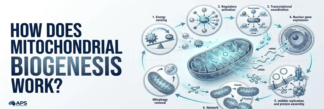
The mitochondrial biogenesis process can be summarised in six connected stages:
- Sensing: Energy stress, calcium flux and other cellular signals activate regulators such as AMPK, p38 MAPK and calcium-sensitive enzymes.
- Transcriptional coordination: PGC-1α coactivates transcription factors including NRF-1, GABP/NRF-2, ERRs and PPARs.
- Nuclear gene expression: The nucleus increases the expression of genes encoding mitochondrial proteins, import machinery and metabolic enzymes.
- mtDNA replication and transcription: TFAM and related proteins help package, maintain, replicate, and transcribe the mitochondrial genome.
- Protein import and assembly: Most mitochondrial proteins are produced in the cytosol, transported through membrane import complexes and assembled within the organelle.
- Network remodelling: Fusion and fission reorganise the mitochondrial network, while mitophagy removes selected damaged components.
These stages overlap rather than following a strictly linear sequence, and each operates on a different timescale. Messenger RNA levels may change within hours of a stimulus, whereas measurable increases in mitochondrial proteins, respiratory capacity or volume generally require repeated stimulation followed by recovery.
The Mitochondrial Biogenesis Pathway: AMPK, SIRT1, PGC-1α and TFAM
No single linear pathway controls every tissue. Instead, several signalling routes converge on a shared regulatory network. A review of the PGC-1 regulatory network describes how PGC-1 coactivators integrate signals involving AMPK, SIRT1, NRFs and ERRα to coordinate mitochondrial gene expression and respiratory function.
| Regulator | What activates or influences it | Main role in the response |
| AMPK | Low cellular energy and a higher AMP-to-ATP ratio | Phosphorylates PGC-1α and shifts metabolism towards ATP-producing processes |
| SIRT1 | NAD+ availability and cellular metabolic state | Deacetylates regulatory proteins, including PGC-1α, in context-dependent signalling |
| p38 MAPK | Muscle contraction and cellular stress | Supports transcriptional responses to exercise and other stressors |
| PGC-1α | AMPK, p38 MAPK, calcium-linked signals and transcriptional feedback | Coactivates multiple nuclear transcription factors |
| NRF1 and related factors | PGC-1α and other transcriptional coactivators | Increase the expression of respiratory, protein-import, and mtDNA-maintenance proteins. |
| TFAM | Nuclear gene expression followed by mitochondrial import | Packages mtDNA and supports its maintenance, transcription, and replication |
This scientific lecture explains how mitochondrial genomes, protein import, growth, and network dynamics work together inside cells.
Why Two Genomes Must Communicate
According to an NCBI overview of mitochondrial genetics, human mtDNA encodes 13 oxidative phosphorylation proteins, 22 transfer RNAs and two ribosomal RNAs. Most proteins needed to build and operate mitochondria are encoded by nuclear DNA.
Functional expansion therefore requires coordination between:
- Nuclear and mitochondrial gene expression
- Protein production and mitochondrial import
- Respiratory-complex assembly
Mitochondria also communicate back to the nucleus. Changes in ATP, calcium, NAD+/NADH balance, reactive oxygen species, and metabolites provide signals about mitochondrial conditions. This process is known as retrograde signalling.
How Fission, Fusion and Mitophagy Fit In
Biogenesis works alongside three mitochondrial quality-control processes. A review of mitochondrial dynamics describes fission and fusion as counterbalancing mechanisms that help maintain mitochondrial distribution, turnover and network quality.
- Fusion mixes mitochondrial membranes and internal components.
- Fission divides the network and can separate damaged regions.
- Mitophagy removes selected damaged or dysfunctional material.
Healthy adaptation depends on balancing the production of new components with network remodelling and the removal of damaged material. Related laboratory investigations may also examine cellular peptides across these interconnected pathways.
Visit Australia Peptide Sciences for more research resources
Why the Response Differs Between Tissues
Mitochondrial content and adaptation vary according to each tissue’s workload, fuel use and specialised function. A stimulus may also produce a local response. Exercise primarily affects working muscles, while cold exposure can strongly influence brown adipose tissue through uncoupling protein 1 (UCP1).
| Tissue | Main metabolic demand | Relevant adaptive feature |
| Heart | Continuous contraction and high ATP turnover | Dense mitochondrial content and strong oxidative capacity |
| Skeletal muscle | Changing demand during movement and exercise | Responsive to repeated contractile activity |
| Liver | Switching between fed and fasted states | Flexible fuel processing and biosynthetic control |
| Brain | Continuous signalling and ion-gradient maintenance | Tight energy regulation and mitochondrial transport |
| Brown adipose tissue | Heat production during cold exposure | UCP1-mediated uncoupling and high mitochondrial content |
Tissue type affects how results are interpreted. A higher mtDNA-to-nuclear DNA ratio may indicate network expansion or changes in cell composition, while whole-tissue oxygen consumption combines multiple cell populations.
Findings must therefore be evaluated according to the tissue, model, stimulus, and sampling time. Results from cultured cells cannot directly represent an intact human organ.
How to Increase Mitochondrial Biogenesis: What the Evidence Supports
Repeated exercise has strong supporting evidence in human skeletal muscle. A scientific review of mitochondrial biogenesis identifies physical exercise as an established physiological stimulus that can activate mitochondrial regulatory pathways.
Muscle contraction alters cellular energy balance, calcium handling, and redox signalling, activating gene transcription and mitochondrial protein synthesis during recovery.
Endurance and interval training provide the clearest evidence for increasing mitochondrial content and respiratory capacity. Resistance exercise may also affect mitochondrial proteins. The response depends on training intensity, duration, total workload, fitness level and the tissue examined.
These exercise-induced adaptations form part of the broader evidence on how to improve mitochondrial function, alongside factors such as nutrition, sleep and metabolic health.
Cold exposure, energy restriction and nutrient signalling have also been studied, but much of the evidence comes from cultured cells or animal models. These findings cannot confirm equivalent effects in humans. Likewise, activating one signalling protein does not prove an increase in mitochondrial content or function.
Exercise-Induced Mitochondrial Biogenesis: From One Session to Training Adaptation
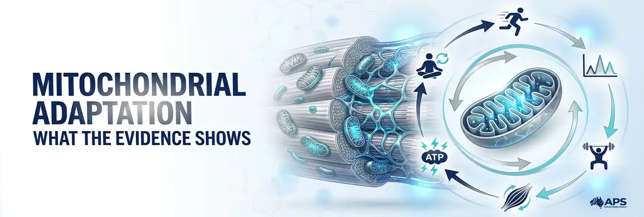
A single exercise session can activate signalling and gene transcription without producing an immediate structural change. During recovery, changes in PGC-1α activity, gene expression and protein synthesis contribute to adaptation. Repeated exercise allows these responses to accumulate.
| Time point | What researchers may observe | What it does not prove alone |
| During or soon after exercise | AMPK or p38 activation, calcium signalling and altered metabolites | A lasting increase in mitochondrial content |
| Hours into recovery | Higher PGC-1α messenger RNA and target-gene transcription | Fully assembled and functional respiratory complexes |
| After repeated training | Increased mitochondrial proteins, enzyme activity, or volume density | Equal adaptation across all tissues or participants |
| After detraining | Partial reversal of training adaptations | Permanent mitochondrial loss or disease |
Training remodels the mitochondrial network by adding new proteins and removing damaged components. The measured response depends on recovery, training load, and baseline fitness.
Mitochondrial Biogenesis and Aging: What Changes Over Time?
Ageing can be associated with reduced respiratory capacity, altered mitochondrial dynamics and accumulated mtDNA damage. However, these changes vary across people and tissues, and physical inactivity may explain part of the difference between younger and older groups.
Older muscle can still adapt to exercise. In a human aerobic-training study, proteins associated with mitochondrial biogenesis, fusion and fission increased in both younger and older men. The response may still vary with training history, health, nutrition, hormonal signalling and programme design, so the pathway does not simply switch off with age.
Measures such as mtDNA copy number, enzyme activity and respiration assess different aspects of mitochondrial quantity and performance. These measurements may also be influenced by the broader biological changes associated with cellular ageing, so they should be interpreted together rather than treated as interchangeable.
Explore Research Peptides in Australia
How Researchers Measure New Mitochondrial Material
There is no single definitive laboratory test for mitochondrial biogenesis. Reliable studies combine molecular, structural and functional measures, interpreted according to the tissue, sampling time and intervention.
| Measure | What it indicates | Important limitation |
| PGC-1α or TFAM RNA/protein | Activation of regulatory pathways | Marker levels may rise before mitochondrial content changes |
| mtDNA-to-nuclear DNA ratio | Relative abundance of the mitochondrial genome | Copy number does not establish respiratory function |
| Citrate synthase activity | Common proxy for mitochondrial content | Enzyme activity may change independently of mitochondrial volume |
| Respiratory-chain proteins | Abundance of selected mitochondrial components | Higher protein levels do not confirm correct assembly or activity |
| Electron microscopy | Mitochondrial volume, density and ultrastructure | Sampling is local and technically demanding |
| Oxygen-consumption assays | Respiratory capacity under defined conditions | Results depend on sample preparation, substrates and normalisation |
| Stable-isotope tracing | Rate of mitochondrial protein synthesis | Requires specialist protocols and does not measure every component |
A review of common mitochondrial research methods shows that morphology, membrane potential, respiration, ATP, ROS and genetic assays answer different questions. MitoTracker staining may also depend on membrane potential, depending on the probe used. No isolated marker can confirm complete mitochondrial formation or improved function.
How to Interpret Mitochondrial Biogenesis Research
No single biomarker confirms that mitochondrial biogenesis has been completed. Researchers therefore interpret molecular signals, mitochondrial content and respiratory function together. A change in one measure can support part of the process without demonstrating that new, fully functional mitochondrial capacity has developed.
| Finding | What it supports | What it does not prove on its own |
| Increased PGC-1α expression or activity | Activation of regulatory pathways associated with mitochondrial biogenesis | Increased mitochondrial content or improved respiratory function |
| Higher mtDNA copy number | Greater abundance of mitochondrial genomes in the sampled cells or tissue | More functional mitochondria or better respiration |
| Higher citrate synthase activity | A commonly used proxy for mitochondrial content and oxidative adaptation in skeletal muscle | Complete mitochondrial formation or improved function across all mitochondrial components |
| Increased oxygen consumption | Greater respiratory activity or capacity under defined experimental conditions | Formation of new mitochondrial material |
These distinctions are important because mitochondrial biogenesis involves coordinated changes in gene regulation, protein synthesis, membrane formation, mtDNA maintenance and respiratory machinery. Stronger evidence therefore comes from combining multiple molecular, structural and functional measurements rather than relying on one marker alone.
Content Versus Function
Mitochondrial content measures how much mitochondrial material is present, while function measures its performance under defined conditions. More material may not increase ATP-linked respiration, and existing mitochondria may become more efficient without increasing in volume.
Mitochondria-targeting compounds such as SS-31 are studied for functional and membrane-related mechanisms. Preclinical research on SS-31 and cardiolipin examined its interaction with inner-membrane structure in a rat ischaemia model; this is distinct from demonstrating the formation of new mitochondrial material.
Common Misunderstandings About Mitochondrial Adaptation
| Misunderstanding | Accurate explanation |
| More mitochondria always means more energy. | ATP output also depends on fuel availability, oxygen, membrane potential, respiratory-complex integrity, and cellular demand. |
| Fission creates new mitochondria by itself. | Fission divides existing material. Network expansion also requires new proteins, lipids, and genetic material. |
| PGC-1α is an on-off switch. | PGC-1α is one coactivator within a broader regulatory network. Its activity alone does not prove completion of the process. |
| One workout builds a new mitochondrial network. | A single session can activate signalling, but lasting structural and functional adaptation usually requires repeated exercise and recovery. |
How Cells Build and Renew Mitochondrial Networks
Cells expand their mitochondrial network by coordinating signalling, gene expression, mtDNA maintenance, protein import, membrane synthesis and quality control. PGC-1α helps organise this response alongside AMPK, SIRT1, NRFs and TFAM.
Exercise provides the best-established human example of mitochondrial biogenesis. However, acute molecular signals do not confirm long-term adaptation. Reliable assessment requires genetic, biochemical, imaging, and functional measurements rather than a single marker.
Frequently Asked Questions About Mitochondrial Biogenesis
What increases mitochondria the most?
Repeated exercise has the strongest human evidence, particularly endurance and interval training, although the response varies by fitness level and training load.
Does walking increase mitochondria?
Regular brisk walking can stimulate mitochondrial adaptation in skeletal muscle, especially in previously inactive people.
What exercise is best for mitochondria?
Endurance and interval exercise have the clearest evidence, while resistance training can provide additional metabolic benefits.
How long does it take to increase mitochondria?
Molecular signals can change within hours, but measurable changes in mitochondrial proteins or respiratory capacity generally require repeated training over several weeks.
Can mitochondria repair themselves?
Cells maintain their mitochondrial network through protein turnover, fusion, fission, and mitophagy, which removes components that are too damaged to repair.
What part of your body has the most mitochondria?
Heart muscle has one of the highest mitochondrial densities because it requires continuous ATP production.
How can I tell if my mitochondria are healthy?
There is no reliable at-home test, so researchers assess mitochondrial health using respiration, enzyme activity, mtDNA, imaging, and other laboratory measurements.
Does CoQ10 increase mitochondria?
CoQ10 supports electron transport and ATP production, but it has not been shown to reliably increase mitochondrial number in healthy humans.
Do mitochondrial supplements really work?
No supplement has been proven to repair or expand mitochondria universally, and results depend on the compound, deficiency, health condition, and evidence available.
What damages mitochondria the most?
No single factor is responsible, but genetic defects, smoking, some toxins, chronic metabolic stress, physical inactivity, and ageing can impair mitochondrial function.
Sources
- PubMed – Vabishchevich et al. 2026
- PubMed – Abrego-Guandique et al. 2025
- PubMed – Mølmen et al.
- AMPK Directly Regulates Muscle PGC-1α
- The Mitochondrial-Derived Peptide MOTS-c Promotes Metabolic Homeostasis
- Metabolic Control of Mitochondrial Biogenesis Through the PGC-1 Regulatory Network
- Primary Mitochondrial Disorders Overview — NCBI GeneReviews
- Mitochondrial Dynamics in Health and Disease
- Mitochondrial Biogenesis: An Update
- Markers of Human Skeletal Muscle Mitochondrial Biogenesis and Quality Control
- Common Mitochondrial Research Methods
- The Mitochondrial-Targeted Compound SS-31 and Cardiolipin