
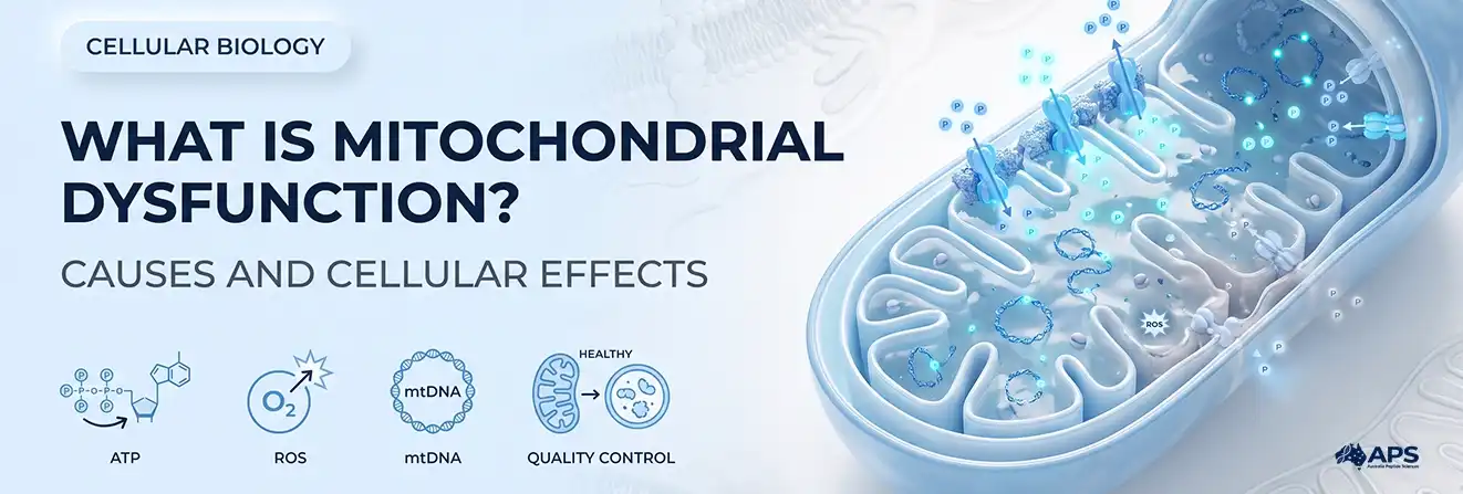
Mitochondrial dysfunction occurs when one or more mitochondrial processes fall below the functional requirements of a specific cell or experimental condition. This may involve inefficient electron transfer, failure to maintain membrane potential, altered redox or calcium signalling, defective genome maintenance, or incomplete removal of damaged organelles. Reduced adenosine triphosphate (ATP) can be one outcome, but it is not the only criterion used to identify mitochondrial impairment.
These changes matter because mitochondria respond differently to nutrient supply, oxygen availability, cellular demand, and experimental conditions. This article follows the sequence from the initiating defect to its measurable cellular consequences, focusing on mechanisms, compensatory responses, and laboratory interpretation rather than lifestyle or nutritional strategies intended to improve mitochondrial performance.
How Researchers Establish a Functional Mitochondrial Baseline
Researchers need a functional baseline before they can determine where mitochondrial failure begins. A separate guide explains normal mitochondrial function more broadly; this section focuses on the processes needed to interpret dysfunction.
NADH and FADH2 donate electrons to the electron transport chain, proton pumping establishes the mitochondrial membrane potential, and ATP synthase uses that gradient during oxidative phosphorylation. The relevant research question is not simply whether ATP is present, but whether respiration, coupling efficiency, reserve capacity, redox balance, and calcium handling remain appropriate for the cell’s demand.
A cell may temporarily preserve cellular energy production by increasing glycolysis, using alternative substrates, or reducing energy use. Therefore, stable whole-cell ATP does not necessarily exclude an early mitochondrial defect.
What Causes Mitochondrial Dysfunction?
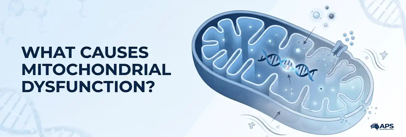
Causes may be genetic, acquired, or arise from interactions between inherited susceptibility, cellular stress, and age-related cellular decline. The initiating event can be difficult to identify because damaged mitochondria may amplify the original disturbance.
1. Genetic Changes in mtDNA or Nuclear DNA
Mitochondrial DNA (mtDNA) encodes 13 proteins required for oxidative phosphorylation, plus RNAs needed for mitochondrial protein synthesis. Most mitochondrial proteins are encoded by nuclear DNA and imported into the organelle.
Pathogenic variants can affect respiratory-chain subunits, protein assembly, mtDNA replication, translation, or fuel metabolism. Some mtDNA mutations are heteroplasmic: mutant and non-mutant genomes coexist in one cell. A defect may appear only above a tissue-specific variant threshold.
2. Acquired and Cellular Stressors
Mitochondrial performance may also be altered by:
- limited oxygen or substrate availability;
- ischaemia followed by reperfusion;
- persistent inflammatory signalling;
- calcium overload;
- metabolic overload and incomplete fuel oxidation;
- damage to mitochondrial proteins or lipids;
- impaired protein folding and removal;
- exposure to particular toxins or medicines; and
- age-related decline in repair and organelle turnover.
Whether a stressor triggers adaptation or damage depends on its dose and duration, the cell type and metabolic context. The experimental endpoint should distinguish a signalling response from impaired mitochondrial function.
What Is the Difference Between Primary and Secondary Mitochondrial Dysfunction?
Researchers classify primary and secondary mitochondrial dysfunction by where the initial defect begins. Primary dysfunction results directly from pathogenic mtDNA or nuclear-DNA variants, while secondary dysfunction develops from another disease, injury, exposure, or cellular disturbance. GeneReviews provides further information on inherited primary mitochondrial disorders.
| Feature | Primary form | Secondary form |
| Initial driver | Pathogenic genetic variant | Another cellular disturbance, disease, or exposure |
| DNA involved | mtDNA or nuclear DNA | No mitochondrial gene variant is required |
| Typical mechanism | Defective respiration, assembly, translation, or mtDNA maintenance | Hypoxia, inflammation, toxicity, metabolic stress, or impaired clearance |
| Course | Often persistent, with variable expression | May be temporary, reversible, or progressive |
| Research question | How does the variant change organelle function? | Is the mitochondrial change a cause, consequence, or amplifier? |
Primary does not always mean maternally inherited. mtDNA is maternally inherited, while nuclear variants may follow autosomal dominant, autosomal recessive, or X-linked patterns. Acquired impairment can still contribute substantially to tissue injury.
How Are Mitochondrial Dysfunction and ATP Production Connected?
ATP output can fall when the respiratory chain cannot transfer electrons efficiently or maintain its proton gradient. Lower membrane potential can also affect protein import, metabolite exchange, and damage signals.
Possible energetic consequences include:
- Less efficient oxidation of NADH and FADH2.
- Reduced proton pumping across the inner membrane.
- Weaker ATP synthase activity.
- A higher reliance on glycolysis.
- Failure of energy-dependent ion pumps when demand exceeds supply.
ATP depletion can disrupt protein synthesis, membrane transport, contraction, and repair. High-demand cells may have limited energetic reserve.
Low respiration does not guarantee a low whole-cell ATP measurement. Cells may increase glycolysis, use alternative fuels, or reduce energy use. A single ATP result may conceal a shift in energy production; combined measurements of respiratory capacity, ATP source, and cellular demand provide a more complete interpretation.
What Is the Connection Between Mitochondrial Dysfunction and Oxidative Stress?
Reactive oxygen species (ROS), including superoxide and hydrogen peroxide, can arise when electrons react prematurely with oxygen. Complexes I and III are important sites, although several enzymes generate ROS.
Controlled ROS signalling supports responses to nutrients, oxygen, and moderate stress. Oxidative stress develops when oxidant generation exceeds antioxidant, repair, and removal capacity.
Under those conditions, oxidation may alter.
- respiratory-chain proteins and metabolic enzymes.
- membrane lipids, including cardiolipin.
- mitochondrial DNA damage and changes to associated proteins.
- ion channels and transporters.
- redox-sensitive signalling pathways.
The relationship is bidirectional: impaired respiration may increase ROS, while oxidative damage can further reduce respiratory efficiency. The effect depends on the amount, location, and duration of ROS, so an increase is not necessarily harmful.
Laboratory studies of antioxidant defence also examine redox-active compounds such as glutathione.
What Are the Major Cellular Effects of Mitochondrial Dysfunction?
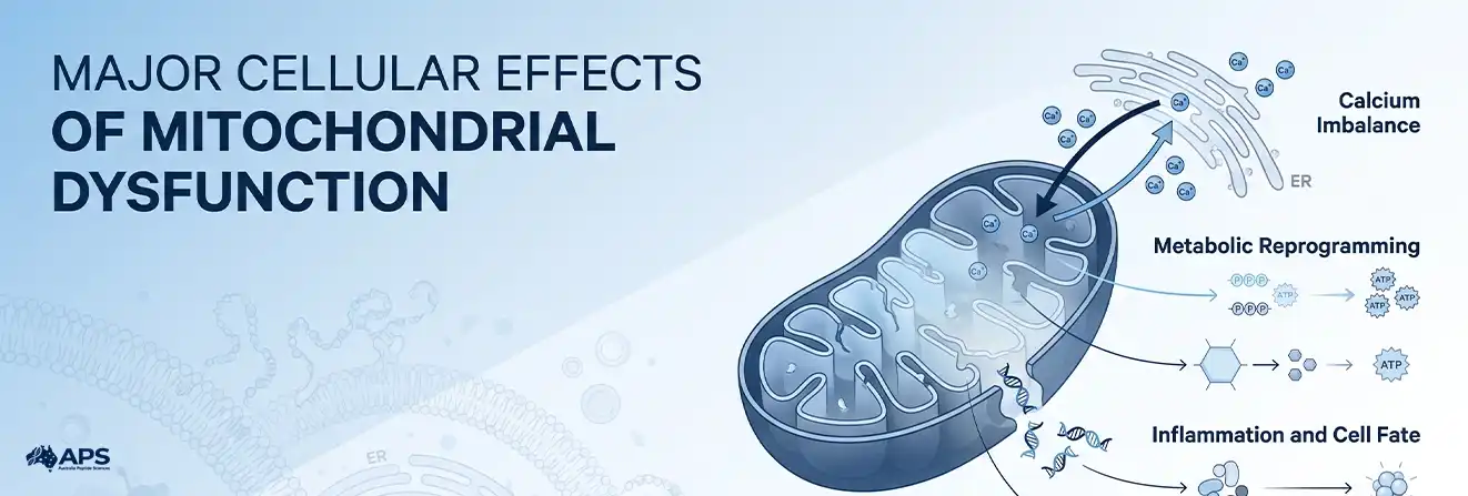
The effects extend across interconnected cellular systems rather than occurring in isolation.
1. Loss of Calcium Control
Mitochondria exchange calcium with the endoplasmic reticulum. Moderate uptake activates matrix enzymes and helps match ATP supply to demand. Excessive uptake, poor efflux, or disrupted contacts can cause overload.
Severe overload may promote permeability transition, depolarisation, swelling, and pro-death signalling. Cytosolic disturbances also affect contraction, secretion, and enzyme activity.
2. Metabolic Reprogramming
Changes in ATP, AMP, NADH, and NAD+ influence energy-sensing pathways such as AMPK, mTOR, and sirtuins. Cells may reduce biosynthesis, increase glycolysis, alter fatty-acid oxidation, or redirect metabolites towards survival pathways.
This response may be protective briefly, but persistent reprogramming can change gene expression, proliferation, and differentiation. Glucose-rich experimental media may hide respiratory defects by supporting glycolysis.
3. Inflammation and Cell Fate
Damaged organelles can expose mtDNA, cardiolipin, or other material as danger signals, potentially activating innate immune and inflammatory pathways.
The eventual outcome depends on damage severity and energetic state:
- mild stress may trigger repair and metabolic adaptation;
- persistent stress may promote cell-cycle arrest or senescence;
- regulated release of cytochrome c can activate apoptosis; and
- profound energy failure and membrane damage may lead to necrotic injury.
The same disturbance may cause adaptation in one cell type and death in another.
The video below discusses mitochondrial disease, cellular energy production and effects across organ systems.
How Does Mitochondrial Quality Control Limit Damage?
Cells maintain mitochondria through repair, remodelling, removal, and replacement. Major components include protein control, mitochondrial fission and fusion, mitophagy, and mitochondrial biogenesis.
Fusion exchanges membranes and contents across the network. Fission distributes organelles and separates poorly functioning regions. Excessive fragmentation or connectivity can both be harmful, so morphology alone cannot establish function.
Mitophagy delivers damaged mitochondria to lysosomes. Loss of membrane potential can initiate PINK1-Parkin or receptor-mediated pathways. An accumulation of autophagic structures may reflect either increased mitochondrial removal or blocked degradation, so it does not by itself establish mitophagy flux.
Mitochondrial biogenesis coordinates nuclear gene expression, protein import, membrane formation and mtDNA replication. PGC-1α, nuclear respiratory factors and TFAM regulate this process, but newly formed mitochondrial components must also be functionally integrated.
Quality control is an integrated system: components may be repaired, mixed through fusion, separated by fission, removed through mitophagy, and replaced through biogenesis.
Why Are Some Cell Types More Vulnerable to Mitochondrial Damage?
Cell vulnerability is determined by the gap between energetic demand and compensatory capacity, not by mitochondrial abundance alone. A tissue may be particularly sensitive when it has high ATP demand, limited glycolytic flexibility, a high proportion of pathogenic mtDNA, weak antioxidant defence, or slow organelle turnover.
Cells carrying the same mtDNA variant may still produce different results because variant load, substrate availability, stress responses, and removal capacity differ between models. These differences are especially relevant to experimental systems used in cellular ageing research.
Researchers should therefore compare matched cell types and report nutrient conditions, oxygen availability, passage number, and normalisation methods before attributing an observed effect to mitochondrial damage.
How Researchers Measure Impaired Mitochondrial Function
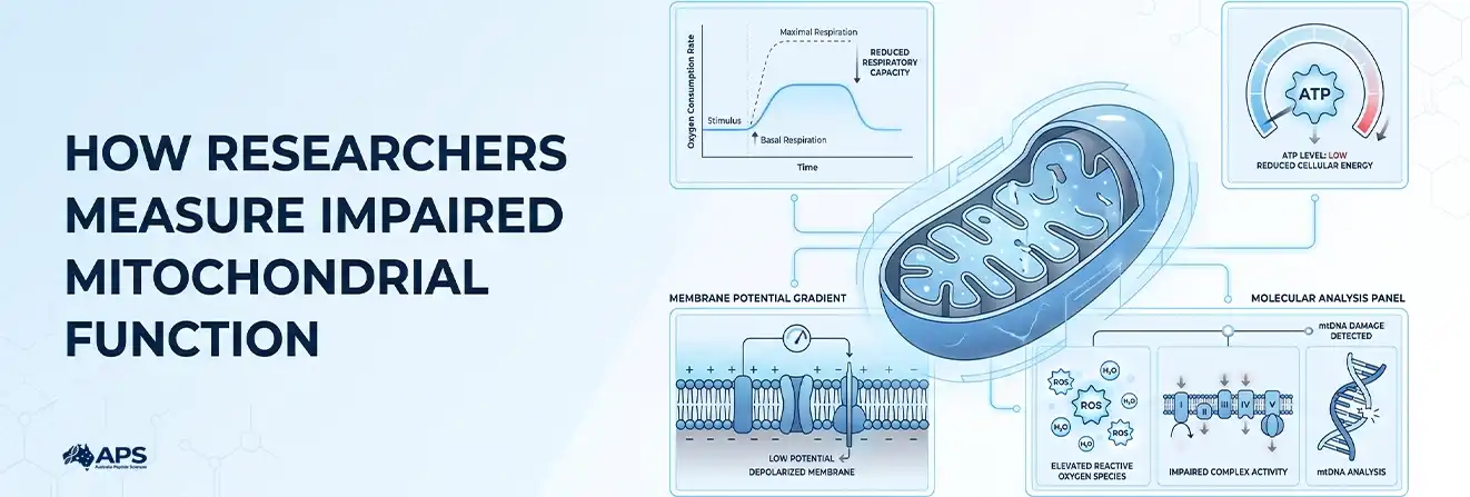
Evidence of mitochondrial dysfunction is strongest when functional, biochemical, and structural measurements agree.
| Measurement | What it assesses | Main limitation |
| Oxygen-consumption rate | Basal respiration, maximal respiration, and reserve capacity | Strongly affected by cell number, substrate and assay conditions |
| ATP assay | Cellular energy status | May not distinguish oxidative ATP from glycolytic ATP |
| Membrane-potential probe | Polarisation of the inner membrane | Dye concentration and mitochondrial mass can affect the signal |
| ROS assay | Changes in oxidant production | Probe specificity and cellular location matter |
| Respiratory-complex assay | Activity of selected electron-transfer complexes | Isolated activity may not reflect intact-cell behaviour |
| mtDNA analysis | Copy number, variants, deletions or lesions | A molecular change does not prove a functional consequence |
| Microscopy | Shape, distribution and network organisation | Fragmentation alone is not a diagnosis |
| Mitophagy-flux assay | Rate of mitochondrial delivery and degradation | Static markers cannot show whether turnover is completed |
Interpretation depends on reported oxygen concentration, nutrient conditions, passage number, tissue origin, and normalisation method.
Standardised oxygen-consumption measurements, combined with other readouts, help distinguish bioenergetic defects from changes in mitochondrial quantity, viability or assay conditions.
How Should Mitochondrial Dysfunction Be Interpreted in Research?
Mitochondrial dysfunction should be interpreted as a pattern across linked measurements, not as a conclusion drawn from one low ATP value, one ROS signal, or one change in mitochondrial morphology. A defensible interpretation connects the initiating defect with changes in respiration, membrane potential, redox state, calcium handling, or quality-control flux.
The strongest studies also distinguish cellular damage from compensation. Stable ATP may reflect increased glycolysis, higher ROS may represent adaptive signalling rather than oxidative injury, and mitochondrial fragmentation may occur without a corresponding loss of respiratory capacity. The cause, cell type, experimental conditions, and measurement timing must therefore be considered together.
Frequently Asked Questions
Over what timescales can mitochondrial quality-control responses occur?
The timescale varies with the experimental model, type and severity of stress, cell type and the relative contributions of repair, mitophagy and mitochondrial biogenesis.
What cellular findings can indicate mitochondrial dysfunction in research?
Research findings may include altered respiration, membrane potential, ATP production, redox balance, calcium handling, mtDNA integrity or mitochondrial quality-control activity. No single measurement is sufficient in every model.
Can mitochondrial dysfunction originate from genetic changes?
Yes. Pathogenic variants in mitochondrial DNA or nuclear genes can disrupt respiratory-chain activity, mitochondrial translation, genome maintenance or related cellular processes.
What damages mitochondria the most?
There is no single greatest cause; severe oxidative stress, pathogenic genetic variants, toxins, oxygen deprivation, inflammation, and calcium overload can all cause substantial damage.
What triggers mitochondrial dysfunction?
It may be triggered by inherited mutations or acquired stresses such as hypoxia, metabolic disruption, inflammation, toxic exposure, and defective mitochondrial quality control.
How is mitochondrial dysfunction assessed experimentally?
Experimental assessment can combine respiration measurements, membrane-potential assays, ATP analysis, ROS measurements, mtDNA analysis, microscopy and mitochondrial quality-control assays.
Can a single biomarker confirm mitochondrial dysfunction?
No. A defensible interpretation generally requires multiple complementary measurements together with the experimental model, cellular context and assay conditions.
Can damaged mitochondria be repaired?
Cells can repair some mitochondrial components, while severely damaged mitochondria may be removed through mitophagy and replaced through mitochondrial biogenesis.
Which organ in the body has the most mitochondria?
The heart has one of the highest mitochondrial densities because cardiac muscle requires a continuous and substantial energy supply.
Can mitochondrial dysfunction occur as both a primary and secondary process?
Yes. Dysfunction may arise directly from mitochondrial or nuclear genetic defects, or secondarily from other cellular stresses, injuries, exposures or disease-associated processes.
References
- Mitochondrial Electron Transport Chain: Oxidative Phosphorylation, Oxidant Production, and Methods of Measurement
- Primary Mitochondrial Disorders Overview
- How Mitochondria Produce Reactive Oxygen Species
- Mitochondrial Quality Control in Human Health and Disease
- A Practical Guide for the Analysis, Standardisation and Interpretation of Oxygen Consumption Measurements