


Retatrutide molecular structure combines a 39-amino-acid peptide chain with selected residue substitutions, a chemical linker and a long fatty-diacid side chain. Known as LY3437943, retatrutide was developed from a glucose-dependent insulinotropic polypeptide (GIP) framework to activate the GIP, GLP-1 and glucagon receptors.
Its design influences receptor activity, resistance to enzymatic breakdown and persistence in circulation.
The complete structure includes more than the amino-acid sequence alone, making it important to distinguish the full lipidated conjugate from database records that describe only the peptide component or calculate its molecular mass using a different structural definition and chemical scope for the recorded molecule.
What Is the Retatrutide Molecular Structure?
Retatrutide is a synthetic, single-chain peptide containing 39 amino-acid residues. Its peptide backbone is derived largely from GIP, but several residues have been altered to balance activity across three related receptors. A lipid-bearing linker attached to a lysine side chain adds another important layer to the design.
The molecule is commonly identified as Retatrutide, LY3437943 or LY-3437943. Its CAS Registry Number is 2381089-83-2, while NOP2Y096GV is its UNII identifier. These identifiers help distinguish the intended molecule from similarly named research materials, salts and records describing related structural forms.
| Property | Description |
| Molecule class | Synthetic, modified peptide conjugate |
| Peptide length | 39 amino-acid residues |
| Development code | LY3437943 |
| CAS number | 2381089-83-2 |
| UNII | NOP2Y096GV |
| Structural origin | GIP-derived peptide framework |
| Main receptor targets | GIPR, GLP-1R and GCGR |
| Key design elements | Non-standard residues, a linker and a long fatty-diacid side chain |
A Single Peptide With Several Structural Layers
The retatrutide peptide molecular structure can be considered in four connected layers. The first is the primary sequence: the order of the 39 residues. The second is the inclusion of non-standard residues that cannot always be represented accurately by an ordinary one-letter amino-acid code. The third is the covalently attached linker and fatty-diacid group. The fourth is the three-dimensional conformation adopted by the molecule in solution or when bound to a receptor.
These layers must be considered together. A sequence written only with standard letters can hide residue modifications, while a molecular formula may represent the full conjugate rather than the peptide chain alone. Likewise, a flat chemical drawing shows connectivity but does not capture the range of shapes available to a large and flexible peptide.
The GIP-Derived Peptide Framework
Retatrutide was engineered from a GIP-based framework rather than assembled by simply joining three natural hormones. The resulting molecule remains one continuous peptide. Selected changes across the chain allow it to interact with the GIP receptor, the GLP-1 receptor and the glucagon receptor.
This distinction matters because the term “triple agonist” describes pharmacological activity, not the number of peptide chains. Retatrutide is not a mixture of three receptor agonists. It is one modified peptide designed to produce agonist activity at three related class B G-protein-coupled receptors.
Retatrutide Chemical Structure Explained
The retatrutide chemical structure contains a conventional peptide backbone made of repeating amide bonds, together with non-standard amino-acid features and a lipid-linked side chain. These modifications change how the molecule behaves without converting it into a small-molecule drug.
Peptide Bonds and Side Chains
Each residue is connected to the next through a peptide bond between an amino group and a carboxyl group. This produces the main backbone, while the chemically varied side chains project outward and influence folding, receptor contact, charge, solubility and enzymatic recognition.
Retatrutide contains acidic, basic, polar and hydrophobic residues. Their arrangement allows parts of the peptide to interact with water and receptor surfaces, while hydrophobic residues contribute to helical structure and receptor-binding contacts. The molecule is electrically and chemically more complex than its elemental formula alone suggests.
The sequence also contains proline- and glycine-rich regions near the C-terminal end. Glycine permits local flexibility, while proline can restrict backbone geometry. Their placement contributes to the overall conformational behaviour of the peptide rather than serving as an interchangeable tail.
Non-Standard Amino-Acid Residues
One important design feature is alpha-aminoisobutyric acid, usually abbreviated Aib. It resembles alanine but carries an additional methyl group on its alpha carbon. This seemingly small change creates steric effects that can reduce recognition by certain peptidases and favour particular backbone conformations.
Retatrutide also includes another methylated residue within its sequence. Such substitutions are important when the molecule is represented in shorthand. Writing a standard amino-acid letter at every position can make the sequence appear natural even when a residue has been chemically modified.
An accurate sequence representation identifies the modified residues explicitly, since these substitutions form part of retatrutide’s chemical identity.
The full chemical identity cannot be recovered reliably from a simplified letter string unless the substitutions, lipid attachment and terminal modifications are also stated.
The Linker and Fatty-Diacid Side Chain
A lysine side chain provides the attachment point for a linker connected to a long fatty diacid. The published chemical design uses a C20 fatty-diacid component rather than the C18 group sometimes attributed to the molecule in secondary summaries. The lipid is not part of the linear 39-residue count, but it is part of the complete covalently bonded molecular structure.
The linker separates the bulky lipid group from the peptide backbone. This spacing helps reduce unwanted interference between the lipid and receptor-binding regions. It also provides the chemical route through which the fatty diacid is joined to the peptide.
The lipid modification promotes reversible association with serum albumin, which can influence clearance and exposure. Urva et al.’s pharmacokinetic study reported a terminal half-life of approximately six days. That measured result cannot be inferred from the structural diagram alone.
C-Terminal Modification
The C-terminal end of retatrutide is amidated rather than left as a free carboxyl group. C-terminal amidation changes the terminal charge and may improve resistance to some forms of exopeptidase activity. It must also be included when calculating the complete elemental composition and mass.
This is another reason that the retatrutide sequence and retatrutide molecular formula should not be treated as independent facts. A sequence may communicate residue order, but the formula depends on every covalent modification and on the precise chemical form selected for calculation.
Retatrutide Sequence and Modified Residues
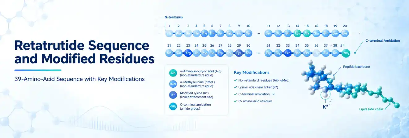
The retatrutide sequence contains 39 residues. A useful shorthand representation is:
Y-Aib-Q-G-T-F-T-S-D-Y-S-I-αMeL-L-D-K-K*-A-Q-Aib-A-F-I-E-Y-L-L-E-G-G-P-S-S-G-A-P-P-P-S-NH2
In this notation, Aib identifies alpha-aminoisobutyric acid, αMeL identifies an alpha-methylated leucine residue, K* marks the lysine bearing the lipid-linker substituent, and NH2 indicates C-terminal amidation. This expanded notation is more informative than a standard one-letter string because it does not conceal the principal modifications.
How to Read the Sequence
The sequence runs from the N-terminus on the left to the C-terminus on the right. The initial tyrosine is position 1, and the amidated serine is position 39. Residues are numbered according to their location in the peptide chain; atoms in the linker and fatty diacid are not counted as additional amino acids.
Accurate representation of the retatrutide sequence requires the linker atoms to remain separate from the 39 amino-acid residues. The non-standard Aib residue, the α-methylated leucine, the lipid-bearing lysine, and the C-terminal amide should also be represented explicitly because each contributes to the defined chemical structure. Omitting these modifications or treating them as standard residues can result in an incomplete representation of the molecule.
Some abbreviated sequences use standard letters and then describe modifications in a separate note. That approach is acceptable only when the notes are complete and clearly connected to the sequence. A string that omits the notes does not fully represent the chemical identity of retatrutide.
Structurally Important Regions
The N-terminal region is especially important for receptor activation. In class B peptide receptors, the peptide’s N-terminus generally enters the receptor’s transmembrane core and contributes to the conformational changes that initiate signalling. Changes in this region can strongly alter potency or receptor preference.
The middle and C-terminal portions help stabilise receptor recognition through contacts with the extracellular domain. Retatrutide’s balanced activity does not arise from one residue acting alone. It results from the combined effect of the GIP-derived scaffold, residue substitutions, side-chain chemistry and the three-dimensional orientation of the peptide at each receptor.
The lipid-bearing lysine performs a different role. Its conjugated side chain primarily supports pharmacokinetic persistence, while the surrounding sequence and linker design help preserve receptor engagement. Structural engineering therefore had to balance prolonged exposure with biological activity.
| Structural feature | Location or form | Main relevance |
| N-terminal region | Beginning of the peptide | Important for receptor activation |
| Aib residues | Modified positions in the chain | Support proteolytic stability and conformational control |
| Alpha-methylated leucine | Internal modified residue | Alters local geometry and enzymatic recognition |
| Lipid-bearing lysine | Internal side chain | Connects the peptide to the linker and fatty diacid |
| C20 fatty diacid | Outside the 39-residue sequence | Promotes reversible albumin association |
| C-terminal amide | End of the peptide | Changes terminal charge and chemical stability |
Retatrutide Molecular Formula and Molecular Weight
The complete chemically defined conjugate commonly associated with retatrutide is reported with the molecular formula C221H342N46O68. Its average molecular weight is approximately 4,731 g/mol.
These values describe a large peptide conjugate and are meaningful only when interpreted alongside the structural definition used for the calculation. PubChem’s record for retatrutide sodium salt provides a reference point for checking the formula, molecular weight and structural form attached to those values.
Understanding the Retatrutide Molecular Formula
The retatrutide molecular formula records the total number of carbon, hydrogen, nitrogen and oxygen atoms in a defined chemical structure. It does not reveal the order of the amino acids, the position of the linker or the shape of the molecule. Two molecules can contain the same elements in the same proportions yet have different connectivity and biological properties.
For a peptide conjugate, the formula depends on several components:
- The elemental composition of all 39 residues after peptide bonds form
- Non-standard residue substitutions
- The lipid-linker substituent
- C-terminal amidation
- The protonation or salt form selected for the record
A structure description and identifier provide essential context for interpreting a reported molecular formula, particularly when different chemical forms or salt states are possible. Quoting only the formula can make it difficult to determine whether the value represents retatrutide itself, a salt, a peptide fragment or another related record.
Understanding the Retatrutide Molecular Weight
The commonly reported retatrutide molecular weight of about 4,731 g/mol is an average molecular mass calculated from the complete structure recorded under the associated chemical entry. Because one dalton per molecule corresponds numerically to one gram per mole, the value may also be expressed as approximately 4,731 Da when discussing a single molecule’s mass.
Average molecular weight is not the same as exact mass or monoisotopic mass. Average molecular weight uses the natural abundance-weighted atomic masses of the elements. Monoisotopic mass assumes a molecule composed of the most abundant isotope of each element. Exact mass is calculated from specified isotopes and a defined structure.
For a molecule as large as retatrutide, these values can differ by several mass units. A laboratory mass spectrum may also show multiple charge states, adducts or counterion-related peaks. The reported mass-to-charge ratio should not be mistaken for the neutral molecule’s molecular weight.
Why Published Formula and Weight Values Differ
Not every database record defines the molecule in the same way. One chemical record may represent the complete lipidated conjugate, while a protein-style registration may calculate an estimated formula from a peptide definition. A salt record may add counterions, and a supplier record may refer to a material containing an unspecified salt, water or residual counterion.
A separate protein-oriented record has reported an estimated formula of C187H281N43O60 and a calculated average mass of 4,091.546 Da.
These values are not directly interchangeable with C221H342N46O68 and approximately 4,731 g/mol because the records use different structural scopes or calculation definitions.
Averaging these values would be inappropriate because they refer to different structural definitions. Their interpretation depends on the specific record, the components included in that definition and the type of mass being reported.
| Structural scope | Reported formula | Reported mass | Appropriate interpretation |
| Full chemically defined conjugate | C221H342N46O68 | About 4,731 g/mol | Use when discussing the complete recorded chemical structure |
| Protein-style registered definition | C187H281N43O60 | 4,091.546 Da, estimated | Keep tied to that specific peptide or protein record |
| Salt or counterion-containing material | Varies | Varies | Specify the salt and composition before comparison |
| Experimental mass-spectrometry result | May not be stated as a formula | Observed m/z values | Interpret charge state, adducts and analytical method |
Two-Dimensional and Three-Dimensional Structure
A two-dimensional chemical drawing and a three-dimensional molecular model answer different questions. Neither should be treated as a complete substitute for the sequence and modification description.
What a 2D Chemical Structure Shows
The 2D structure maps covalent connectivity. It can show the peptide bonds, residue side chains, stereochemistry, linker, lipid group and terminal amide in one diagram. It is the most direct way to establish which atoms are connected.
However, a full 2D depiction of retatrutide is visually dense. The molecule contains hundreds of heavy atoms and many stereochemical centres. A high-resolution diagram is therefore necessary, ideally accompanied by an annotated simplified view that identifies the peptide chain, modified residues, linker and lipid side chain.
Long IUPAC names, SMILES strings and InChI records encode aspects of the same connectivity for database and software use. They are valuable for machine processing but are not practical replacements for an explanatory figure. Copying a long identifier into body text adds little value unless the reader needs it for a specific cheminformatics task.
Why There Is No Single Simple 3D Shape
Retatrutide is large and flexible, with many rotatable bonds. In solution, it does not remain locked in one rigid conformation. Its shape can change with solvent conditions, temperature, concentration, albumin association and receptor binding.
This flexibility also makes ordinary small-molecule conformer generation difficult. A rendered 3D model may display one energetically plausible pose, but it represents only one of several possible conformations. Such models depend on the starting geometry, force field and assumptions used during calculation.
Receptor-bound structural studies answer a more focused question. They can show how the peptide is positioned when interacting with GIPR, GLP-1R or GCGR. In these complexes, the receptor helps stabilise a particular peptide conformation, often with a substantial helical region and an N-terminal segment extending into the transmembrane binding pocket.
Primary, Secondary and Receptor-Bound Structure
The term “molecular structure” can refer to several levels:
- Primary structure:the order and chemical identity of the residues.
- Covalent structure:the peptide plus all linkers, lipid groups and terminal modifications.
- Secondary structure:local patterns such as an alpha helix.
- Solution conformation:the changing set of shapes populated in a particular environment.
- Receptor-bound structure:the conformation stabilised during interaction with a target receptor.
Clear labelling prevents a sequence diagram, 2D chemical drawing and receptor complex from being presented as though they show the same type of information.
How the Structure Supports Triple-Receptor Agonism
Within GLP-1 peptide research, retatrutide is examined as a single engineered peptide with activity at GIPR, GLP-1R and GCGR.
These three targets are related class B GPCRs that recognise peptide hormones using an extracellular domain and a transmembrane binding pocket.
The video below explains retatrutide’s triple-agonist mechanism.
Two-Part Receptor Engagement
Peptide agonists in this receptor family generally follow a two-part interaction model. The C-terminal portion of the peptide first contributes to recognition by the extracellular domain. The N-terminal portion then engages the transmembrane core, stabilising the active receptor conformation required for intracellular signalling.
Retatrutide’s sequence must satisfy this process at three receptors whose natural ligands are related but not identical. Its GIP-derived backbone provides a starting framework, while selected substitutions adjust the pattern of charge, hydrophobicity, steric bulk and hydrogen-bonding potential along the chain.
The result is not equal binding through exactly the same contacts at every receptor. Each receptor presents a distinct binding environment. Retatrutide can adopt compatible interactions across the three targets, but its relative potency and signalling profile are determined experimentally rather than from the sequence alone.
Structure Does Not Equal Clinical Effect
The molecular design explains how triple agonism is possible, but it does not establish clinical efficacy or safety. Receptor assays, experimental models and clinical studies answer different questions and should not be treated as interchangeable evidence.
These levels of evidence should remain separate. A structural feature may support a mechanistic hypothesis, but measured pharmacokinetic and biological outcomes require direct experimental evidence under defined conditions. The actual half-life, for example, must be established experimentally rather than assumed from molecular design.
Retatrutide and Tirzepatide Structural Differences
Retatrutide and tirzepatide are both single-chain, lipidated peptide agonists derived from GIP-related design principles. Their structures include non-standard residues and albumin-binding lipid modifications, but their receptor profiles are different.
Tirzepatide is designed for dual GIPR and GLP-1R agonism. Retatrutide adds meaningful glucagon-receptor agonism, making it a triple agonist. This distinction reflects changes across the peptide sequence rather than the addition of a third peptide component.
| Feature | Retatrutide | Tirzepatide |
| Peptide form | Single modified peptide | Single modified peptide |
| Peptide length | 39 residues | 39 residues |
| Receptor profile | GIPR, GLP-1R and GCGR | GIPR and GLP-1R |
| Backbone design | GIP-derived | GIP-derived |
| Stability strategy | Non-standard residues and lipidation | Non-standard residues and lipidation |
| Lipid-bearing design | C20 fatty-diacid component through a linker | Different linker and attachment design |
| Main structural distinction | Sequence balanced for triple agonism | Sequence balanced for dual agonism |
Structural differences between retatrutide, tirzepatide and mazdutide extend beyond fatty-acid chain length or a single attachment position. Related molecular and mechanistic comparisons are compiled in the Peptide Comparison Guides.
Receptor selectivity arises from the entire peptide-receptor interface. Differences at the N-terminus, internal residues, C-terminal recognition region and lipid-linker placement can all affect activity and exposure.
How to Interpret a Retatrutide Structure Record
Chemical and biological databases often organise peptides differently. A careful record check prevents most formula, sequence and molecular-weight errors.
Confirming Molecular Identity
Accurate identification begins with the primary name and associated identifiers. Retatrutide is associated with LY3437943, CAS 2381089-83-2 and UNII NOP2Y096GV. A matching name alone may be insufficient when a record refers to a sodium salt, an alternative structural definition or a supplier-specific preparation.
The interpretation of a structure record depends on whether it includes all 39 amino-acid residues, the non-standard residues, the substituted lysine, the complete linker, the fatty-diacid side chain, C-terminal amidation and any counterion, salt or solvate.
Differences in these components can produce legitimate differences in the reported molecular formula and mass, provided that the values are labelled according to their structural scope.
Mass records may report average molecular weight, calculated mass, exact mass, monoisotopic mass or observed mass-to-charge ratio. These represent different quantities, so meaningful comparison requires equivalent chemical definitions and mass types.
Separate Computed Data From Experimental Results
A database can calculate formula, exact mass, hydrogen-bond counts and other descriptors from a submitted structure. Those values describe the structure as entered. They do not confirm the identity, purity or composition of a physical sample.
Experimental characterisation may use mass spectrometry, chromatography and peptide mapping. Each method answers a different analytical question. A commercial purity percentage, for example, does not establish that the sequence and full conjugate are correct unless identity has also been verified with suitable methods.
The Research Peptide Guides place molecular structure alongside analytical quality and laboratory handling considerations.
Research and Regulatory Context in Australia
Retatrutide remains an investigational molecule and should not be presented as an established therapeutic product in Australia. A description of its molecular structure does not establish approval, clinical suitability or permission for personal use.
Clinical trial registries document ongoing investigational studies of retatrutide, but registry listings do not establish therapeutic approval or suitability for personal use.
In this context, research-use terminology refers specifically to materials supplied for controlled analytical or experimental work rather than human consumption. Batch-level identity, purity and composition should be supported by appropriate analytical documentation such as a Peptide Certificate of Analysis (COA).
Australian regulatory status can change over time, with current therapeutic approvals recorded in the Australian Register of Therapeutic Goods.
Structural information describes the molecule itself and does not establish prescribing, dosing or administration guidance.
The retatrutide research compound is supplied for controlled laboratory research and is not represented for human or veterinary use.
Retatrutide combines a modified GIP-derived peptide backbone with lipidation and terminal modifications that contribute to its structural and pharmacological properties.
Together, these elements support stability, prolonged exposure and agonist activity at GIPR, GLP-1R and GCGR. The complete chemically defined conjugate is commonly recorded as C221H342N46O68 with an average molecular weight of about 4,731 g/mol.
Different database values should not be combined without checking whether they describe the full conjugate, a peptide-style definition, a salt or another structural form. Sequence, formula, 2D connectivity and receptor-bound conformation each describe a different aspect of the same engineered molecule.
FAQs About Retatrutide Molecular Structure
What Exactly Is Retatrutide?
Retatrutide is an investigational 39-amino-acid synthetic peptide engineered to activate the GIP, GLP-1 and glucagon receptors. Its structure includes modified residues and a lipid-linked side chain designed to improve stability and extend circulation time.
How Is Retatrutide Different From Semaglutide?
Semaglutide activates the GLP-1 receptor, while retatrutide is designed to activate the GIP, GLP-1 and glucagon receptors. Retatrutide is also a distinct 39-residue peptide with its own sequence and lipid-linked structure.
Is Tirzepatide a Peptide or a Small Molecule?
Tirzepatide is a synthetic 39-amino-acid peptide, not a small molecule. It is engineered as a dual agonist of the GIP and GLP-1 receptors and includes structural modifications that support prolonged activity.
Sources
https://pubchem.ncbi.nlm.nih.gov/compound/171934787
https://pubmed.ncbi.nlm.nih.gov/35985340/
https://pubmed.ncbi.nlm.nih.gov/36354040/