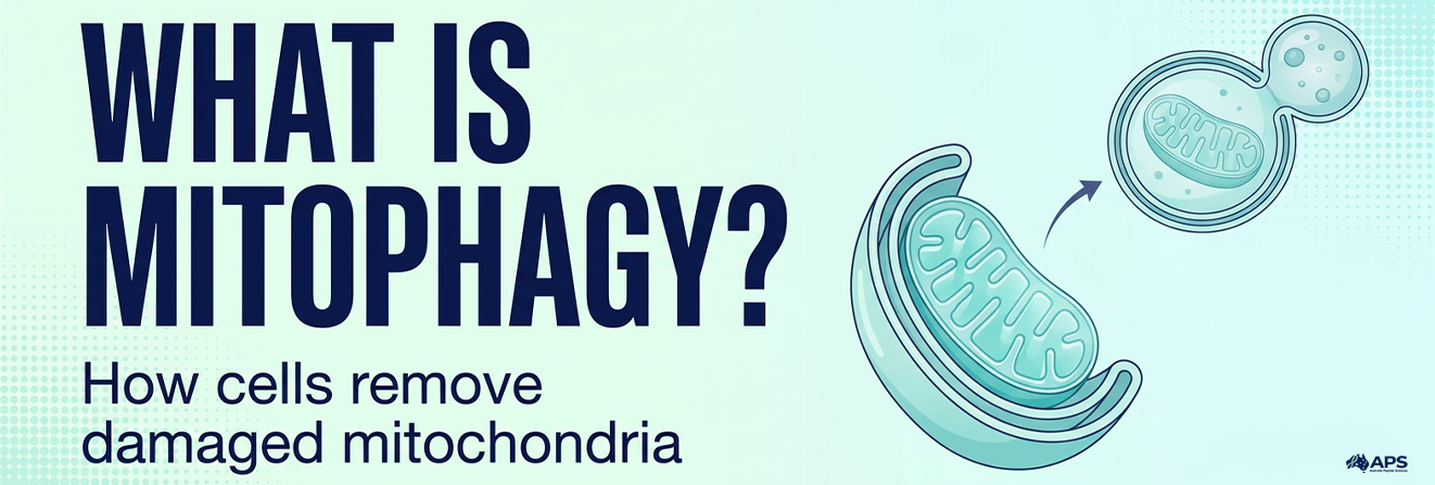
Mitophagy is the selective breakdown of mitochondria through the cell’s autophagy-lysosome system, helping remove damaged or unnecessary mitochondria and maintain cellular function.
Mitochondria support energy production, metabolism and cellular signalling. Cells maintain them by repairing damage where possible and removing mitochondria that are no longer needed.
Several pathways control this process, allowing cells to respond to mitochondrial damage, changing metabolic demands and developmental signals. Understanding these pathways helps explain how cells coordinate mitochondrial removal with renewal.
Research-use notice: This article is provided for scientific and educational purposes only. It does not constitute medical advice, treatment guidance or instructions for the human or veterinary use of research compounds. Findings from cell and animal studies should not be interpreted as established clinical benefits.
Quick Answer
Mitophagy delivers selected mitochondria to lysosomes for degradation. It supports mitochondrial quality control through mechanisms including PINK1-Parkin signalling and receptor-mediated pathways. Removal signals, autophagosome formation, and lysosomal breakdown must work together. The biological outcome depends on the tissue, the cellular conditions, and whether clearance is completed.
What Is Mitophagy, and Why Do Cells Need It?
A practical mitophagy definition is the selective autophagic degradation of mitochondria. The term mitochondrial autophagy refers to the same process and distinguishes it from pathways that remove other cellular material.
Mitochondria exchange components, divide and adjust their activity as cellular demands change. Studies involving GHK-Cu or other compounds must distinguish effects on cellular signalling from evidence of mitochondrial clearance. Damage to mitochondrial proteins, membranes and DNA creates a need for maintenance, but demonstrating their removal requires measurements of the clearance process itself.
Mitophagy is part of mitochondrial quality control, alongside protein maintenance and changes to the mitochondrial network. It allows cells to remove selected mitochondria when they are damaged or no longer needed.
Cells also remove mitochondria during normal development. During red blood cell maturation, for example, mitochondrial clearance helps cells acquire their mature form. Experimental work on erythroid maturation identified an important role for the mitochondrial protein NIX in this process.
Mitophagy therefore supports routine turnover and development as well as responses to damage.
Mitophagy vs Autophagy: What Is the Difference?
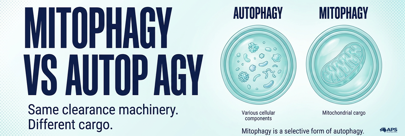
When comparing mitophagy vs autophagy, the key distinction is the material targeted. Autophagy encompasses a broad group of cellular degradation processes; mitophagy specifically targets mitochondria.
In macroautophagy, a membrane surrounds selected cellular material and delivers it to lysosomes for breakdown. Mitophagy uses this machinery together with signals that identify mitochondrial cargo.
| Feature | Autophagy | Mitophagy |
| Material targeted | Can include proteins and various organelles | Mitochondria |
| Selectivity | Can be selective or non-selective | Requires mitochondrial recognition |
| Research interpretation | A general autophagy measurement does not establish mitochondrial clearance | Requires evidence of mitochondrial targeting and turnover |
“Cellular recycling” is a useful shorthand, but it has limits. Mitophagy does not directly turn a damaged mitochondrion into a new one. Its breakdown products can re-enter cellular metabolism, while mitochondrial biogenesis helps renew the mitochondrial population.
How Does Mitophagy Work?
Mitophagy involves mitochondrial recognition, recruitment of autophagy machinery, enclosure of selected material and lysosomal degradation. In the typical macroautophagic pathway, selected mitochondria are enclosed within an autophagosome before being delivered to lysosomes.
1. Mitochondria Receive Signals for Removal
Changes in mitochondrial condition or cellular requirements influence which mitochondria are selected.
One signal is the loss of mitochondrial membrane potential, the electrical component of the gradient across the inner membrane that supports production of adenosine triphosphate (ATP), a molecule cells use to transfer energy. Other signals can arise during low oxygen availability, metabolic adaptation or cellular development.
Different conditions activate different recognition mechanisms. No single mitophagy pathway explains every form of mitochondrial turnover.
2. Receptors Recruit Autophagy Machinery
Molecular tags and receptors connect selected mitochondria to the machinery that forms the surrounding membrane.
Some mechanisms use ubiquitin, a small protein attached to other proteins as a cellular signal. Others depend on receptors embedded in mitochondrial membranes.
These interactions help determine where the isolation membrane, known as the phagophore, forms around the mitochondrial cargo.
3. The Cargo Is Enclosed
The phagophore expands around the selected material and closes to form a double-membrane autophagosome.
Remodelling of the mitochondrial network can help separate material intended for degradation. However, fragmentation alone does not prove that mitophagy has occurred because fragmented mitochondria may remain in the cell without reaching lysosomes.
4. Lysosomes Degrade the Material
The autophagosome fuses with a lysosome, where enzymes break down the enclosed mitochondrial material.
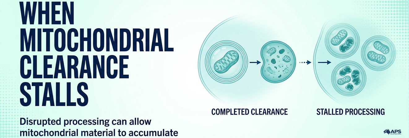
Successful damaged mitochondria removal requires recognition, enclosure and effective lysosomal degradation. If downstream processing is impaired, mitochondrial cargo can accumulate despite strong removal signals.
The PINK1 Parkin Pathway: Recognising Mitochondrial Damage
The PINK1 Parkin pathway is a well-studied mechanism that links mitochondrial damage signals to selective clearance.
PINK1 is a protein kinase that modifies other molecules by adding phosphate groups. Parkin, encoded by the PRKN gene, is an E3 ubiquitin ligase that helps attach ubiquitin to target proteins.
1. PINK1 Accumulates When Mitochondrial Import Is Disrupted
In healthy, polarised mitochondria, PINK1 is continuously imported and processed, keeping its abundance low. When substantial loss of membrane potential disrupts this process:
- PINK1 accumulates on the outer mitochondrial membrane.
- PINK1 phosphorylates ubiquitin.
- The resulting signal helps recruit and activate Parkin.
2. Parkin Amplifies the Removal Signal
Activated Parkin strengthens the mitochondrial removal signal through several connected steps:
- Parkin adds ubiquitin chains to proteins on the outer mitochondrial membrane.
- These chains recruit autophagy receptors, particularly OPTN and NDP52.
- The receptors connect the marked mitochondrion to proteins involved in forming the enclosing membrane.
- Autophagy machinery then prepares the selected mitochondrial cargo for lysosomal degradation.
A mechanistic study identified OPTN and NDP52 as key receptors in this process.
Parkin helps mark and organise mitochondrial cargo for removal; it does not degrade mitochondria itself.
This pathway is particularly informative in experimental models of mitochondrial depolarisation. However, its prominence in laboratory studies does not establish that Parkin is required for every form of mitochondrial turnover.
For a visual explanation of mitochondrial damage recognition, watch this PINK1-Parkin video from Cell Press.
Parkin-Independent Mitophagy: Other Routes to Clearance
Parkin-independent mitophagy includes several mechanisms that can promote mitochondrial clearance without requiring Parkin.
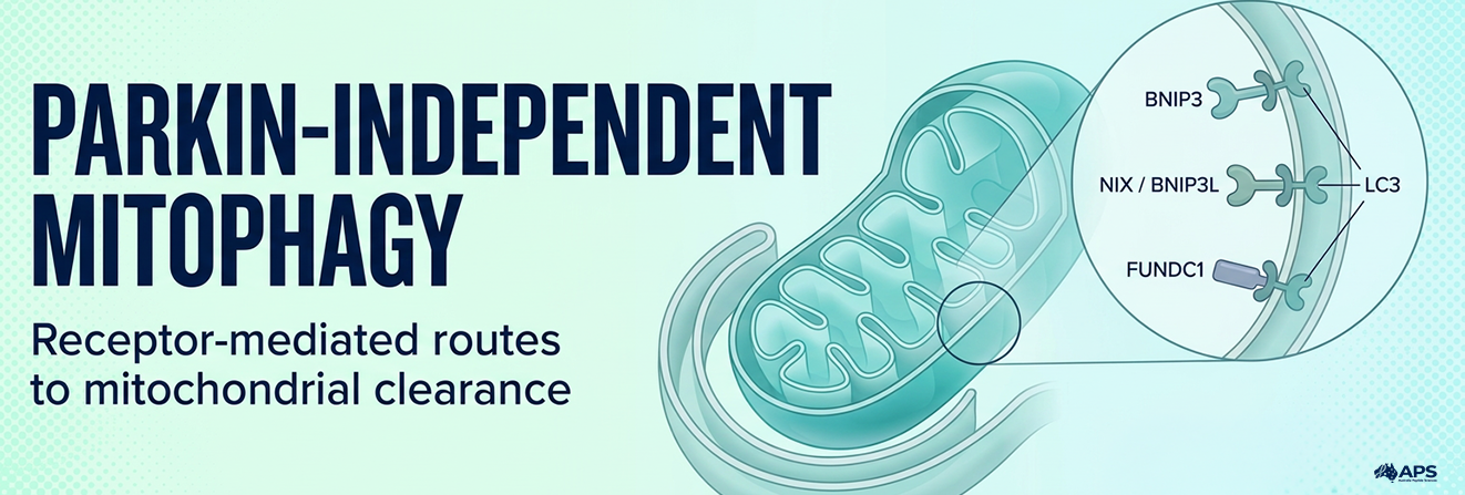
Some mitochondrial membrane proteins act as receptors that connect selected mitochondria to autophagy-associated proteins such as LC3.
| Receptor | Research context | Relevant function |
| BNIP3 | Hypoxia and other stress responses | Connects mitochondria with autophagy machinery |
| NIX/BNIP3L | Developmental clearance and stress responses | Supports mitochondrial removal, including during erythroid maturation |
| FUNDC1 | Hypoxia-related mitophagy | Interacts with LC3 through a regulated binding region |
In the original FUNDC1 study, disrupting its interaction with LC3 impaired hypoxia-induced mitophagy in mammalian cells. This finding supports a receptor-mediated route to mitochondrial clearance.
The terminology requires care. “Parkin-independent” does not automatically mean “PINK1-independent” or “ubiquitin-independent,” because these terms describe different molecular requirements.
Animal studies also show why biological context matters. Research using a mito-QC mouse reporter found that basal mitophagy persisted without PINK1 in several metabolically active tissues. These findings indicate that cells can maintain mitochondrial turnover through multiple context-dependent mechanisms.
A 2026 review in Nature Reviews Molecular Cell Biology examines PINK1-Parkin-dependent and independent mitophagy pathways, highlighting how their regulation and physiological roles vary across mammalian tissues. Despite substantial mechanistic progress, the conditions governing these pathways in living organisms remain incompletely understood.
What Triggers Mitophagy, and How Is It Regulated?
Common mitophagy triggers include mitochondrial damage, altered oxygen availability, metabolic changes and developmental signals. Their effects depend on the cell type, biological context and machinery available to respond.
1. Damage, Oxygen and Metabolic Signals
Different cellular conditions can activate distinct clearance mechanisms:
- Loss of membrane potential can initiate PINK1-associated signalling.
- Hypoxia can activate receptor-mediated mechanisms.
- Changes in nutrient availability can affect the wider autophagy system.
- Developmental signals can remove mitochondria that specialised cells no longer require.
Reactive oxygen species participate in cellular signalling, while oxidative stress can contribute to molecular damage when redox balance is disrupted. Neither finding alone establishes that mitochondria have been selected and successfully degraded.
In metabolic health research, mitochondrial turnover is considered alongside energy production and nutrient use. Measuring clearance alone therefore provides only part of the picture of cellular metabolic function.
2. Regulation Extends Beyond Initiation
Mitophagy regulation involves energy and nutrient sensors, receptor modifications, autophagosome formation and lysosomal capacity.
Several proteins help coordinate these stages:
- AMPK responds to changes in cellular energy availability and can promote autophagy-associated signalling.
- mTORC1 integrates nutrient and growth signals and can suppress autophagy initiation when active.
- ULK1 helps initiate the formation of autophagic membranes.
These proteins regulate early stages of the pathway, while successful turnover also depends on lysosomal capacity. If downstream degradation is impaired, stronger initiation signals may leave more mitochondrial material awaiting breakdown.
Experimental triggers should not be translated directly into universal fasting durations, exercise schedules or personal protocols.
Mitophagy and Mitochondrial Biogenesis: Balancing Removal and Renewal
The relationship between mitophagy and mitochondrial biogenesis helps cells maintain a mitochondrial population suited to their functional needs.
Mitophagy removes selected mitochondria. Biogenesis produces mitochondrial components, imports proteins and coordinates nuclear and mitochondrial gene activity to support growth and renewal.
Fusion and fission also help organise the mitochondrial network. Fusion allows mitochondria to exchange contents, while fission can separate sections of the network for redistribution or removal.
Research in Caenorhabditis elegans demonstrated coordination between removal and biogenesis, linking these processes with mitochondrial content and longevity in the worm model. This finding does not establish an equivalent effect on human lifespan.
In mitochondrial research involving compounds such as SS-31, researchers need to distinguish changes in mitochondrial abundance from changes in function. Measuring energy production alongside turnover helps assess whether the mitochondrial population can meet the cell’s metabolic demands.
Related NAD+ research examines how cellular energy metabolism interacts with mitochondrial biology. Changes in NAD+ metabolism and mitophagic clearance are distinct outcomes, so evidence for one should not be used as a substitute for measuring the other.
Explore research peptides at Australia Peptide Sciences
What Happens When Mitochondrial Clearance Is Disrupted?
Impaired clearance can allow dysfunctional mitochondrial material to accumulate. Depending on the biological context, this may affect energy production, cellular stress responses and inflammatory signalling.
However, mitochondrial dysfunction and defective mitophagy are not interchangeable terms. Mitochondria can perform poorly because of respiratory-chain defects, genetic changes or other disturbances, while the clearance pathway may be active, insufficient or blocked at a specific stage.
A mitochondrial muscle disease study illustrates this complexity.
Researchers observed increased mitophagy during earlier disease stages and stalled processing in more advanced affected muscle fibres. The study examined both mouse models and human muscle samples.
A single tissue sample may therefore contain regions with different clearance states. Measurements averaged across the entire tissue can obscure this variation.
Disease associations also require careful interpretation. Finding altered mitophagy in a disorder does not establish that increasing it will treat the condition. Researchers must determine where the defect occurs, whether it contributes to the disease and what happens when the pathway is modified.
Mitophagy and Aging: What Does Longevity Research Show?
Research on mitophagy and aging examines how mitochondrial maintenance changes over time and affects tissue function within the wider biology of cellular aging.
These changes vary across tissues and experimental models.
1. Animal Findings and Human Outcomes Answer Different Questions
Studies of mitophagy and longevity link mitochondrial maintenance with lifespan in model organisms, but do not establish equivalent effects in humans.
Human studies usually assess physical performance, metabolic measures and molecular biomarkers.
| Study type | What it can establish | Main limitation |
| Cell experiment | Molecular mechanisms under controlled conditions | Does not reproduce the interactions of a whole organism |
| Animal study | Tissue effects and, in some models, lifespan outcomes | Findings may not translate directly to humans |
| Human observational study | Associations between measured variables | Cannot establish causation on its own |
| Randomised human trial | Effects on predefined outcomes in the studied population | Does not establish unmeasured mechanisms or lifespan effects |
2. What Human Compound Studies Actually Measure
Urolithin A, a non-peptide metabolite, provides an example of why evidence must be assessed for each compound separately. Its findings in mitochondrial and ageing research cannot establish the effects of a different research compound, including a peptide such as KPV.
A randomised trial in older adults reported improvements in selected muscle-endurance measures at two months and changes in plasma biomarkers at four months. However, the between-group differences in the primary outcomes, six-minute walking distance and maximal ATP production in hand muscle, were not statistically significant at four months.
These findings apply to the outcomes measured in that study. They do not demonstrate human lifespan extension or directly quantify completed mitophagy throughout the body.
When reviewing materials in the Cellular Research Peptides category, check which model and outcomes were studied for each compound. Products grouped in the same category may have different mechanisms and levels of evidence; research-use-only materials should not be treated as established therapies.
How Do Researchers Measure Mitophagy?
Researchers assess mitophagy by tracking mitochondrial cargo and determining whether it reaches the degradation system. The main challenge is distinguishing pathway initiation from completed turnover.
1. Fluorescent Reporters Track Lysosomal Delivery
Reporters such as mito-QC and mt-Keima use fluorescence changes to detect mitochondrial material entering acidic compartments.
The mito-QC methodology combines mitochondrial targeting with green and red fluorescent signals. Inside acidic lysosomes, the green signal is quenched while the red signal remains detectable, helping researchers identify delivered mitochondrial material.
These reporters provide more specific information than mitochondrial abundance alone. However, their interpretation still depends on reporter behaviour, imaging conditions and lysosomal function.
2. Mitophagic Flux Requires More Than a Snapshot
Mitophagic flux describes progression through the pathway over time.
Several observations are insufficient on their own:
- Increased PINK1 or Parkin may indicate mitochondrial recognition or signalling.
- More LC3-associated structures may reflect increased formation or reduced turnover.
- Lower mitochondrial mass may result from reduced biogenesis, cell injury or other processes.
- Mitochondrial-lysosomal overlap does not establish the rate of completed degradation.
A stronger assessment combines measurements of mitochondrial targeting, lysosomal delivery and turnover with suitable controls. Time-course data can distinguish temporary responses from sustained changes.
Controls must also match the assay. In pH-sensitive reporters, interventions that change lysosomal acidity can alter fluorescence directly and complicate interpretation.
When evaluating a study, separate evidence of completed clearance from evidence of improved mitochondrial function, and check whether the experimental model supports the authors’ conclusions.
Why Balanced Mitochondrial Turnover Matters
Mitophagy helps cells manage their mitochondrial population through selective removal and degradation. Effective turnover depends on completing and coordinating the entire process, rather than activating a single protein.
Research findings depend on the pathway, tissue, model and measurements used. Distinguishing mitochondrial targeting from completed degradation and improved function keeps conclusions about human health and longevity grounded in what each study actually demonstrates.
Discover Australia Peptide Sciences and explore our research resources
Frequently Asked Questions About Mitophagy
Is mitophagy a form of cell death?
No; mitophagy removes mitochondrial material within a cell and is distinct from processes that destroy the entire cell.
Does mitophagy happen only when mitochondria are damaged?
No; it also supports routine mitochondrial turnover and programmed removal during cellular development.
Is mitochondrial fission the same as mitophagy?
No; fission divides mitochondria, whereas mitophagy targets mitochondrial material for lysosomal degradation.
Does every cell use the same mitophagy pathway?
No; pathway dependence varies with cell type, developmental stage, metabolic conditions and the stimulus involved.
Can mitophagy happen without Parkin?
Yes; receptor-mediated and other mechanisms can support mitochondrial clearance without requiring Parkin.
Does increased LC3 prove that mitophagy has increased?
No; LC3 measurements alone do not establish mitochondrial targeting or completed lysosomal degradation.
How long does fasting take to activate mitophagy?
There is no established universal fasting duration that reliably activates mitophagy across human tissues.
Is more mitophagy always beneficial?
No; its effects depend on cellular needs, successful degradation and coordination with mitochondrial renewal.
Is urolithin A a peptide?
No; urolithin A is a non-peptide metabolite produced by certain gut microbes from dietary precursors.
Does increasing mitophagy extend human lifespan?
Human lifespan extension through targeted increases in mitophagy has not been established.
References
- Lazarou M, et al. The ubiquitin kinase PINK1 recruits autophagy receptors to induce mitophagy. Nature. 2015.
- McWilliams TG, et al. Basal Mitophagy Occurs Independently of PINK1 in Mouse Tissues of High Metabolic Demand. Cell Metabolism. 2018.
- Liu L, et al. Mitochondrial outer-membrane protein FUNDC1 mediates hypoxia-induced mitophagy in mammalian cells. Nature Cell Biology. 2012.
- Chen M, et al. Selective mitochondrial autophagy during erythroid maturation. Autophagy. 2008.
- Palikaras K, Lionaki E, Tavernarakis N. Coordination of mitophagy and mitochondrial biogenesis during ageing in C. elegans. Nature. 2015.
- Mito T, et al. Mosaic dysfunction of mitophagy in mitochondrial muscle disease. Cell Metabolism. 2022.
- Liu S, et al. Effect of Urolithin A Supplementation on Muscle Endurance and Mitochondrial Health in Older Adults: A Randomized Clinical Trial. JAMA Network Open. 2022.
- Montava-Garriga L, et al. Semi-automated quantitation of mitophagy in cells and tissues. Mechanisms of Ageing and Development. 2020.